A Synchronous Presentation of Two Different Ovarian Tumors: A Rare Occurrence
- *Corresponding Author:
- Dr. Divya Sethi
Department of Pathology, C-5/20, Sector-11, Rohini, Delhi, India.
E-mail: dr.divyasethi@gmail.com
Abstract
Approximately 60% of all ovarian tumors are epithelial in origin, and these neoplasms are thought to arise from the ovarian surface epithelium or small epithelial inclusion cysts. Surface epithelium is capable of differentiating into serous (tubal), mucinous, endometrioid or transitional epithelium. Serous and mucinous cystadenomas are the most common epithelial tumors and, together, account for about 30% of ovarian tumors We report a case of a 29‑year‑old lady P1L1 presenting with the chief complaints of pain abdomen off and on since the last 1 year. Ultrasonography revealed normal uterus with enlarged right ovary, with a cyst measuring 46 mm × 36 mm × 55 mm showing internal echoes with volume of 50 cc., left ovary also enlarged with multiple well‑defined cysts measuring 34 mm × 44 mm × 69 mm with volume of 55 cc and the largest cyst measuring 37 mm. Bilateral ovarian cystectomy was done and sent for histopathology. To our surprise, both the ovaries revealed different histopathological pictures, with the right ovary revealing serous cystadenoma and the left ovary showing mucinous cystadenoma. This rare occurrence has never been reported so far in the literature to the best of our knowledge.
Keywords
Bilateral ovarian tumors, Serous and mucinous cystadenomas, Synchronous ovarian tumors
Introduction
Approximately 60% of all ovarian tumors are epithelial in origin.[1,2] Except for rare mucinous tumors developing from teratomas, these are thought to arise from the ovarian surface epithelium or small epithelial inclusion cysts.[3,4] Because the surface epithelium of the ovaries is derived from the coelomic epithelium, which also gives rise to the Müllerian ducts, it is accepted that the surface epithelium is capable of differentiating into serous (tubal), mucinous, endometrioid or transitional epithelium. Serous and mucinous cystadenomas are the most common epithelial tumors and, together, account for about 30% of ovarian tumors. Occurrence of mixed epithelial tumors is rare, but the presence of serous cystadenoma in one ovary and mucinous cystadenoma in the other has never been reported in the literature and this is the first one to be reported to the best of our knowledge.
Case Report
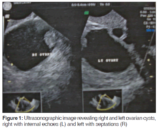
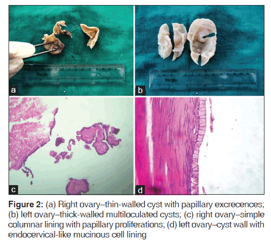
A 29-year-old lady P1L1 presented to the gynecology outpatient department with chief complaints of pain abdomen off and on since the last 1 year. The pain was predominantly in the right iliac fossa region. She kept taking medication for the pain until recently in January 2012, when she developed acute pain also on the left side of the abdomen for which she was admitted. She also complained of one episode of vomiting. Ectopic pregnancy was ruled out by urine pregnancy test. Abdominal examination did not reveal any mass and no abnormality was detected on per speculum examination. Bimanual examination revealed a normal-sized uterus and a cystic mass felt laterally near the posterior fornix that was approximately 4 cm in diameter. An ultrasound examination and CA-125 were requested. Ultrasonography revealed normal uterus with enlarged right ovary with a cyst measuring 46 mm × 36 mm × 55 mm showing internal echoes with volume of 50 cc, the left ovary was also enlarged with multiple well-defined cysts measuring 34 mm × 44 mm × 69 mm with volume of 55 cc and the largest cyst measured 37 mm [Figure 1]. The serum CA-125 level was 38 U/mL. Diagnostic laparoscopy followed by laparotomy with bilateral ovarian cystectomy was performed, followed by chromopertubation under general anesthesia. Intraoperative findings–adhesions present with omental and fat adhesions with the abdominal wall. The right cyst was approximately 5 cm in diameter with smooth surface and surface papillary excrecences and containing straw-colored fluid. The left ovarian cyst was approximately 5 cm × 4 cm, with a bosselated surface. Cut-surface revealed multiloculated cyst with thick walls. Omentum was healthy. On chromopertubation, bilateral tubes were patent. Bilateral ovarian cystectomy specimens were received in the Department of Pathology. On gross examination, the right ovary revealed an opened up cystic structure approximately 6 cm in diameter. The inner surface of the wall revealed multiple papillary excrescences. The left ovary was 7 cm in diameter, with the cut surface revealing multiloculations with solid and cystic areas. Microscopic examination of the right ovary revealed cyst wall lined by simple columnar lining with papillary proliferations at places, and the left cyst revealed a thick-walled cyst with endocervical like mucinous cell lining [Figure 2a-d]. Thus, a diagnosis of right ovarian serous cystadenoma with left ovarian mucinous cystadenoma was made.
Discussion
A female’s risk at birth of having ovarian tumor sometime in her life is 6-7%. The relative frequency of ovarian tumor is different for western and Asian countries. Two-third of ovarian tumors occur in women of the reproductive age group.[5]
Other studies also show that most ovarian tumors occur in women of the reproductive age group. The peak incidence of ovarian tumor is between 21 and 40 years. Benign ovarian tumors occur in all age groups, whereas malignant ovarian tumors are more common in the elderly.[6] Majority of the benign serous tumors occur in the 4th-6th decades, although they may occur in patients younger than 20 years or older than 80 years.[7] Mucinous cystadenoma may occur at any age, but is most often diagnosed in the 4th-6th decades. Mucinous cancers have a mean age of 53-54 years.[8]
Of the three types, epithelial tumors are the most common, comprising about 58% of all ovarian tumors. Serous and mucinous cystadenomas are the most common epithelial tumors and, together, account for about 30% of ovarian tumors. Serous tumors constitute about 30% of all ovarian tumors, making them the single most common group of epithelial tumors. They comprise 22% of benign and nearly 50% of malignant primary tumors of the ovary. Of all serous tumors, 50% are benign, 15% are borderline and 35% are invasive carcinomas.[9,10]
About 20% of benign serous tumors are bilateral. Microscopically, benign serous tumors are lined by ciliated and non-ciliated low columnar cells with bland ovoid basal nuclei.
There are small foci of mild to moderate nuclear atypia or nuclear stratification in occasional, otherwise benign, serous tumors. When these features are present in only a few low-power fields (>5-10% of the tumor), the clinical evolution is invariably benign.
Mucinous cystadenoma is the most common ovarian mucinous tumor; it is about equal in incidence to serous cystadenoma. Borderline and malignant mucinous tumors are less numerous than their serous counterparts, and borderline mucinous tumors outnumber mucinous carcinomas. Mucinous cystadenoma is generally unilateral. The cut surface reveals unilocular or multilocular mucin-filled cysts of varying size. Microscopically, a layer of columnar cells lines the cysts, papillae and crypt-like structures that are found in benign mucinous tumors. A majority of the tumor cells are endocervical-like or gastric-like, with uniform round or oval basal nuclei and clear or amphophilic cytoplasm.[11]
The simultaneous occurrence of multiple primary cancers in the upper female genital tract is well known, but most of them are malignant in nature.[12] In the present case, however, the presence of serous cystadenoma in one ovary and mucinous cystadenoma in the other was observed.
Conclusion
There have been reports with simultaneous occurrence of two different tumors at different sites in the female genital tract; however, there has not been a single case reported with one-sided ovary revealing serous cystadenoma and the other side showing mucinous cystadenoma, as in the present case.
Source of Support
Nil.
Conflict of Interest
None declared.
References
- Katsube Y, Berg JW, Silverberg SG. Epidemiologic pathology of ovarian tumors: A histopathologic review of primary ovarian neoplasms diagnosed in the Denver Standard Metropolitan Statistical Area, 1 July-31 December 1969 and 1 July-31 December 1979. Int J Gynecol Pathol 1982;1:3-16.
- Koonings PP, Campbell K, Mishell DR Jr, Grimes DA. Relative frequency of primary ovarian neoplasms: A 10-year review. Obstet Gynecol 1989;74:921-6.
- Powell DE, Puls L, van Nagell J Jr. Current concepts in epithelial ovarian tumors: Does benign to malignant transformation occur? Hum Pathol 1992;23:846-7.
- Scully RE, Young RH, Clement PB. Tumors of the Ovary, Maldeveloped Gonads, Fallopian Tube, and Broad Ligament. Washington, DC: Armed Forces Institute of Pathology; 1998. Atlas of Tumor Pathology; Third series, Fascicle 23.
- Jha R, Karki S. Histological pattern of ovarian tumors and their age distribution. Nepal Med Coll J 2008;10:81-5.
- Merino MJ, Jaffe G. Age contrast in ovarian pathology. Cancer 1993;71:537-44.
- Tavassoli FA, Devilee P. WHO Classification of Tumors. Pathology and Genetics, Tumors of Breast and Female Genital Organs. Lyon, France: IARC Press; 2003.
- Scully Robert E, Young Robert H, Clement Phillip B. Atlas of Tumor Pathology. Tumors of the ovary, maldeveloped gonads, fallopian tube and broad ligament. 3rd series, Fascicle 23. AFIP, 1999
- Russell P. The pathological assessment of ovarian neoplasms. Introduction to the common “epithelial” tumours and analysis of benign ‘epithelial’ tumours. Pathology 1979;11:5-26.
- Purola E. Serous papillary ovarian tumors. A study of 233 cases with special reference to the histological type of tumor and its influence in prognosis. Acta Obstet Gynecol Scand 1963;42:1-77.
- Tenti P, Aguzzi A, Riva C, Usellini L, Zappatore R, Bara J, et al. Ovarian mucinous tumors frequently express markers of gastric, intestinal, and pancreatobiliary epithelial cells. Cancer 1992; 69:2131-42.
- Guiu XM. Simultaneous carcinomas involving the endometrium and ovaries. Advances in surgical pathology of the ovary 1999;32:287-8.


 The Annals of Medical and Health Sciences Research is a monthly multidisciplinary medical journal.