
Assessment of the Applicability of KPG Index and Grisar's Method of Three Dimensional Evaluation of Canine Position in Cases with Unilateral Cleft Lip and Palate: An Observational Prospective Study: A Study Protocol
Received: 14-Sep-2022, Manuscript No. AMHSR-22-74639; Editor assigned: 16-Sep-2022, Pre QC No. AMHSR-22-74639 (PQ); Reviewed: 30-Sep-2022 QC No. AMHSR-22-74639; Revised: 28-Jan-2023, Manuscript No. AMHSR-22-74639 (R); Published: 08-Feb-2023, DOI: 10.54608.annalsmedical.2023.84
Citation: Lakhe P, et al. Assessment of the Applicability of KPG Index and Grisar's Method of Three Dimensional Evaluation of Canine Position in Cases with Unilateral Cleft Lip and Palate: An Observational Prospective Study- A Study Protocol. Ann Med Health Sci Res. 2023;13:442-445
This open-access article is distributed under the terms of the Creative Commons Attribution Non-Commercial License (CC BY-NC) (http://creativecommons.org/licenses/by-nc/4.0/), which permits reuse, distribution and reproduction of the article, provided that the original work is properly cited and the reuse is restricted to noncommercial purposes. For commercial reuse, contact reprints@pulsus.com
Abstract
Background: Dental problems are among the most serious complications a patient having a cleft lip and palate may face. Impacted canine are 10-20 times more common than the overall population. Treatment for impacted maxillary canines typically requires surgery and is laden with risk. Therefore, it is desirable to have a reliable method for early canine position recognition.
Aim and objectives: To Study the effectiveness o f t he KPG index and Grisar's method of three-dimensional evaluation position of canine in cases with "unilateral cleft lip and palate" is the goal of this observational prospective study.
Methodology: After receiving the patient's permission, a total of 15 cone beam computed tomography pictures will be taken. Each patient's canine position will be assessed in the frontal, sagittal, and lateral views.
Result: The end result is that conventional radiography techniques might make it challenging to diagnose and plan therapy due to the overlap of structures on the film. The goal of this study is to determine if the previously published method can be used to aid clinicians in rapidly approximating the difficulty of tr eating impacted canines without requiring the clinician to take numerous measurements before informing the cleft lip and palate patient of the approximate treatment plan.
Conclusion: The ability to precisely locate impacted canines through the use of spatial relationships and high quality tissue contrast is made possible by 3D volumetric imaging systems employing CBCT images. In order to better plan therapy, orthodontists need information about the exact location and orientation variation of the impacted canines.
Keywords
Diagnosis; Treatment planning; Panaromic radiograph; Missing
Introduction
One of the most frequent congenital abnormalities affecting the face and skull is a cleft lip or palate. They account for around one in seven hundred births worldwide [1,2]. Asia is the region with the greatest cleft prevalence rate, with one cleft birth for every 500 inhabitants. People with clefts are more likely to have irregularly shaped, sized, and positioned teeth [3,4]. Cleft patients have a considerably greater rate of impaction of canine when compared to the overall population [4]. The rate reaches 82% in patients having Clefting of Lip and/or Palate (UCLP) on one side only [5-7]. In the general population, the number of people who have it ranges from 0.9% to 2.2% [8,9]. This difference in frequency could be due to the unique features of the cleft, agenesis, and extra teeth in the cleft area, as well as the surgeries done in that area [10]. Canine eruptive disorders are visible on panoramic radiographs as an abnormally large mesial crown tilt toward the midline [11,12] or as an overlap of the cusp and root of the permanent maxillary lateral incisor. [13-15]. Grisar K et al. evaluated the location of canines using Cone Beam Computed Tomography (CBCT) images and gave a 3D cataloguing of localizing the position of canines. In 2009, Kau CH, et al. came up with a new method called the KPG Index to help clinicians quickly estimate how hard it will be to treat impacted canines using CBCT scans. Panoramic xrays are two-dimensional images that don't show the buccolingual positioning of impacted canines or how the roots of nearby teeth have worn down. By their very nature, CBCT images are much better than 2D images in many ways. When a 3D view is available, it is easy to answer many diagnostic questions about impacted canines. Older ways of looking at impacted teeth have been replaced by CBCT. This technique helps doctors find the right diagnosis and plan the right treatment for impacted canines by locating the impaction in all three dimensions. In the near future, the KPG index and Grisars method will be very important to orthodontics. This is because 3D imaging is becoming more and more popular, and people with cleft lip and palate often have impacted canines. Patients and parents want to know how long treatment will take. This index should help an orthodontist estimate how long it will take to treat these impactions [16-18]. There isn't much written about how three-dimensional classification systems of canine impaction can be used to figure out where the maxillary canine in individual of unilateral cleft lip and palate. So, the goal of this observational prospective study research is to figure out whether or not the KPG index and Grisar's method useful for evaluating the position of a canine in three dimensions in individuals having "unilateral cleft lip and palate.
Research question PICOT format
Are KPG index and Grisar’s method of three dimensional evaluation of canine position applicable in cases with unilateral cleft lip and palate?
• PICOT: ‘Cleft lip and palate cases’.
• Assess the applicability of KPG index and Grisar’s method of three dimensional evaluation of canine position in cases with UCLP 1 year.
The purpose of this prospective observational study is to determine whether the KPG index and Grisar's threedimensional evaluation of canine position are useful in diagnosing and treating UCLP.
• To assess ‘the applicability of KPG index for evaluation of canine position in cases with UCLP using three-dimensional cone beam CT.
• To assess the ‘applicability of Grisar’s classification for evaluation of canine position in cases with UCLP using three-dimensional cone beam CT.
Literature Review
This prospective observational study will be conducted in the department of orthodontics and will be completed in one year after receiving approval by the IREC. A total of 15 patients who report to the department's OPD and are registered with Smile Train will be chosen for the study. Written consent will be acquired from the guardian of each case. The patients in the study will be UCLP patients who meet the following exclusion and inclusion criteria.
The inclusion criteria consist of the non-syndromic patients having UCLP and patient in the ‘aged between (9-13) years. The patient that will be excluded from the study were patient with missing canine, syndromic cases, cases with bilateral cleft lip and palate, cases with ABG, cases with ‘isolated cleft palate and uvula’, cases with cleft alveolus, unilateral/ bilateral cleft lip, deciduous dentition. For the sample size calculation formula will be applied [19].
Sample size calculation
Formula for sample size with acceptable margin error: Daniel formula for sample size estimation
Where;
At 5%, the degree of significance is 2/2.
In other words, the 95 percent confidence interval is 1.96.
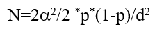
P=prevalence=2.2%=0.022
D=desired error of margin=7%=0.07
N =1.962 × 0.01 × (1 - 0.01)/0.052=15 cases required.
• 95% confidence interval.
• One-sided nature of the test.
• Statistical test: Descriptive statistics and frequency distribution.
• Formula reference: Daniel, et al.
Methodology
For the sample, cone beam computed tomography images will be obtained. Planmeca romexis software will be used to section the images (slice thickness 3 mm. The position of canine will be assessed using KPG index and Grisars method.
For each case, parameters measured will be as follows:
• Parameters taken from the KPG index.
• Position of canine in the anterior-posterior direction in the transverse "X" axis.
• Position of canine in the "superior-inferior direction on the "vertical Y" axis".
• Position of canine in the transverse dimension along the z axis.
• Parameters taken from the Grisars method.
• Canine position in the the mesiodistal region.
• Canine position in the bucco-palatal region.
• Canine position in the transverse dimension.
• Parameters taken from the KPG index.
• Canine cusp tip on the X-axis in the anteroposterior direction.
• First, no intervention is required if the root tip of the canine cusp has erupted correctly.
• Canine cusp and root apexes are located on either side of the midline of the alveolar ridge.
• The third dimension is the distance from the edge of the alveolus to the tip of the cusp or the end of the root of the adjacent tooth, which may be the distall. Half of the lateral incisor or the mesial half of the first bicuspid.
• Fourth, the opposing tooth's cusp or root is located in the lingual half of the lateral incisor or lingual half of the first bicuspid.
• Fifth, the central incisor's cusp or root tip is located behind the first premolar and in advance of the second premolar's midline.
• Canine cuspal tip in the superior inferior direction of the y axis.
• The tip of the cusp of the canine is in the right superior inferior position.
• The tip of the cusp of the canine is in the crown area.
• The tip of the cusp of the canine is in line with the cervical 1/3rd of the root of the incisor.
• The tip of the cusp of the canine is in line with the middle 1/3rd of the root of the incisor.
• The tip of the cusp of the canine is in line with the apical 1/3rd of the root of the incisor.
• The tip of the cusp of the canine is above the root of the incisor.
• Canine root tip in the vertical direction on the y axis.
• The vertical position of the canine cusp/root tip is correct.
• The incisor root's apical 1/3rd third is on a horizontal plane with the root tip.
• The position of apical end if root of the incisor root is in a horizontal plane in the middle 1/3rd of the root.
• The root tip of the incisor tooth is parallel to the cervical 1/3rd.
• The root tip is in the crown region.
• The root tip includes the area past the crown.
• Position of the cuspal tip of canine on the Z-axis in the transverse dimension.
• The canine cusp/root tip is in the right spot along the occlusal arch.
• The tip of the cusp or the tip of the root is between 0 and 2 mm from the side of the tooth that comes into contact with the other teeth.
• The tip of the cusp or root is 2.0-4.0 mm from the occlusal side of the alveolar ridge.
• The tip of the cusp or root is four to six millimetres (mm) from the side of the alveolar ridge opposite the occlusal arch.
• From the occlusal arch, the tip of the cusp or root is set between 6.0 and 8.0 mm away.
• The cusp or root tip of the opposite canine is more than 8.0 mm away from the occlusal side of the alveolar ridge, either buccally or palatally.
• Parameters taken from the Grisars method.
Mesio-distal positioning of the canine as related to neighboring teeth:
• Mesialy oriented crown and distaly oriented root apex.
• Distally oriented crown and mesialy oriented root apex.
• Vertically positioned.
• Horizontally positioned.
• Ectopically position.
The vertical position of the maxillary canine's cusp in relation to the adjacent tooth:
• Cervical one third.
• Middle one third.
• Apical one third.
• Supra-apical.
Canine position in the bucco-palatal region:
• Vestibular region.
• Intra-alveolar region.
• Palatal region.
Statistical test:
• SPSS22. Version.
• Descriptive statistics and frequency distribution.
Discussion
For effective treatment of impacted canines, it is important to get an accurate diagnosis, and a CBCT image can show where it is in space. Because of its benefits, a CBCT image is the best way to use radiography to find both the impaction and the structures around it. It is hoped that a clinician could use the KPG index and Grisar's method to get a good idea of how long it would take to bring that tooth into the mouth.
Implication
Compared to a group of patients without clefts, those with complete alveolar clefts have a very different position of their canines during eruption and are more likely to have their canines get stuck. There have been reports that canine impaction makes orthodontic treatment take longer, makes the mechanics of treatment harder, and raises the cost of treatment. So, the best way to deal with maxillary canines that are stuck is to find them early and stop them from becoming stuck. CBCT scans and threedimensional (3D) volumetric imaging systems can be used to figure out where the affected canines are by looking at how they are arranged in space and how different the tissues look from each other. Orthodontists can make better treatment plans if they know the exact location and range of orientation of the impacted canines.
Conclusion
The ability to precisely locate impacted canines through the use of spatial relationships and high quality tissue contrast is made possible by 3D volumetric imaging systems employing CBCT images. In order to better plan therapy, orthodontists need information about the exact location and orientation variation of the impacted canines.
References
- Shapira Y, Lubit E, Kuftinec MM. Congenitally missing second premolars in cleft lip and cleft palate children. Am J Orthod Dentofacial Orthop. 1999;115:396-400. [Crossref] [Google Scholar] [PubMed]
- Vyas T, Gupta P, Kumar S, Gupta R, Gupta T, Singh HP. Cleft of lip and palate: A review. J Family Med Prim Care. 2020;9:2621. [Crossref] [Google Scholar] [PubMed]
- Beaty TH, Murray JC, Marazita ML, Munger RG, Ruczinski I, Hetmanski JB, et al. A genome-wide association study of cleft lip with and without cleft palate identifies risk variants near MAFB and ABCA4. Nat Genet. 2010;42:525-529. [Crossref] [Google Scholar] [PubMed]
- Troxell JB, Fonseca RJ, Osbon DB. A retrospective study of alveolar cleft grafting. J Oral Maxillofac Surg. 1982;40:721-725. [Crossref] [Google Scholar] [PubMed]
- El Deeb M, Messer LB, Lehnert MW, Hebda TW, Waite de. Canine eruption into grafted bone in maxillary alveolar cleft defects. Cleft Palate J. 1982;19:9-16. [Google Scholar] [PubMed]
- Mangione F, Nguyen L, Foumou N, Bocquet E, Dursun E. Cleft palate with/without cleft lip in French children: radiographic evaluation of prevalence, location and coexistence of dental anomalies inside and outside cleft region. Clin Oral Investig. 2018;22:689-695. [Crossref] [Google Scholar] [PubMed]
- Celikoglu M, Buyuk SK, Sekerci AE, Cantekin K, Candirli C. Maxillary dental anomalies in patients with cleft lip and palate: a cone beam computed tomography study. J Clin Pediatr Dent. 2015;39:183-186. [Crossref] [Google Scholar] [PubMed]
- Dachi SF, Howell FV. A survey of 3,874 routine full-mouth radiographs. I. A study of retained roots and teeth. Oral Surg Oral Med Oral Patho. 1961;14:916–924. [Crossref] [Google Scholar] [PubMed]
- Thilander B, Myrberg N. The prevalence of malocclusion in Swedish schoolchildren. Scand J Dent Res. 1973;81:12-21. [Crossref] [Google Scholar] [PubMed]
- Ericson S, Kurol J. Radiographic examination of ectopically erupting maxillary canines. Am J Orthod Dentofacial Orthop. 1987;91:483-492. [Crossref] [Google Scholar] [PubMed]
- Bishara SE. Impacted maxillary canines: A review. Am J Orthod Dentofacial Orthop. 1992;101:159-171. [Crossref] [Google Scholar] [PubMed]
- Dewinter G, Quirynen M, Heidbuchel K, Verdonck A, Willems G, Carels C. Dental abnormalities, bone graft quality, and periodontal conditions in patients with unilateral cleft lip and palate at different phases of orthodontic treatment. Cleft Palate Craniofac J. 2003;40:343-50. [Crossref] [Google Scholar] [PubMed]
- Power SM, Short MB. An investigation into the response of palatally displaced canines to the removal of deciduous canines and an assessment of factors contributing to favourable eruption. Br J Orthod. 1993;20:215-23. [Crossref] [Google Scholar] [PubMed]
- Ericson S, Kurol J. Radiographic examination of ectopically erupting maxillary canines. Am J Orthod Dentofacial Orthop. 1987;91:483-492. [Crossref] [Google Scholar] [PubMed]
- Russell KA, Mcleod CE. Canine eruption in patients with complete cleft lip and palate. Cleft Palate Craniofac J. 2008;45:73-80. [Crossref] [Google Scholar] [PubMed]
- Jung YH, Liang H, Benson BW, Flint DJ, Cho BH. The assessment of impacted maxillary canine position with panoramic radiography and cone beam CT. Dentomaxillofac Radiol. 2012;41:356-60. [Crossref] [Google Scholar] [PubMed]
- Grisar K, Piccart F, Al-Rimawi AS, Basso I, Politis C, Jacobs R. Three-dimensional position of impacted maxillary canines: Prevalence, associated pathology and introduction to a new classification system. Clin Exp Dent Res. 2019;5:19-25. [Crossref] [Google Scholar] [PubMed]
- Kau CH, Pan P, Gallerano RL, English JD. A novel 3D classification system for canine impactions-the KPG index. Int J Med Robot. 2009;5:291-6. [Crossref] [Google Scholar] [PubMed]
- Daniel WW, Cross CL. Biostatistics: A foundation for analysis in the health sciences. John Wiley and Sons, 11th ed, New York, United States, 2018. [Google Scholar]


 The Annals of Medical and Health Sciences Research is a monthly multidisciplinary medical journal.