Effect of Kinesthetic Illusion Induced by Visual Stimulation on Hand Function in Patients with Chronic Stroke: A Pilot Randomized Controlled Trial
Received: 29-Nov-2023, Manuscript No. AMHSR-23-121605; Editor assigned: 01-Dec-2023, Pre QC No. AMHSR-23-121605 (PQ); Reviewed: 15-Dec-2024 QC No. AMHSR-23-121605 (Q); Revised: 26-Dec-2024, Manuscript No. AMHSR-23-121605 (R); Published: 03-Jan-2025
Citation: Vanu VB, et al. Effect of Kinesthetic Illusion Induced by Visual Stimulation on Hand Function in Patients with Chronic Stroke: A Pilot Randomized Controlled Trial. Ann Med Health Sci Res. 2024;14:1-11
This open-access article is distributed under the terms of the Creative Commons Attribution Non-Commercial License (CC BY-NC) (http://creativecommons.org/licenses/by-nc/4.0/), which permits reuse, distribution and reproduction of the article, provided that the original work is properly cited and the reuse is restricted to noncommercial purposes. For commercial reuse, contact reprints@pulsus.com
Abstract
Background: Upper extremity motor dysfunction is the major problem in stroke patients which leads to dependency during ADL and reduces their QOL. Rehabilitation of hand function is the major challenge seen in neuro rehabilitation settings. Kinesthetic Illusion Induced by Visual Stimulation (KINVIS) is an implicit motor imagery that provides kinesthetic and visual stimulus to the patients as they visualize their own body part moving in the video. This provides a visual illusion to the patient. Participants: 30 participants with chronic stroke of age 45 to 65 were included in the study. Methods: 30 participants with chronic stroke were allocated into two groups i.e. experimental group and control group. Experimental group underwent KINVIS along with conventional therapy for 4 weeks while control group underwent conventional physiotherapy alone for 4 weeks. Outcomes measures used in the study to assess the hand function were fugl meyer assessment for upper extremity, box and block test and modified ashworth scale. Analysis: Statistical analysis of data was done using SPSS 27.0 version software. Analysis was done using Kolmogorov Smirnov test, descriptive and inferential statistics using chi square test, student’s paired and unpaired t test. Results: The results of the study within group comparison showed significant improvement in both experimental group and control group for FMA and BBT after 4 weeks of intervention. (p-value 0.001) Between group analyses for FMA and BBT was significant with experimental group showing more statistically significant improvement as compared to the conventional group. The KINVIS group demonstrated notable improvement in wrist flexor muscle tone according to the Modified Ashworth Scale (MAS). Conclusion: KINVIS is found to be effective to improve hand function in patients with chronic stroke.
Keywords
KINVIS; Chronic stroke; Hand function; Rehabilitation
Introduction
Stroke (cerebrovascular accident) is the “sudden loss of neurological function caused by an interruption of blood flow to the brain” [1]. It is a leading cause of premature death and disability in economically developing countries [2,3]. The most common residue of the stroke is hemiparesis and tonal abnormalities which lead to motor impairments affecting person’s daily life and quality of living. Reports showed that up to 85% of stroke survivor’s experiences unilateral weakness of upper limb and lower limb and that 55% to 75% continued to have impairments in upper-extremity and hand functions [4,5]. The negative outcome of not regaining functional use of the arm after stroke is documented to occur in a range of one-third to two-third of cases, and only small percentage of patients i.e. around 5% to 20% of patients with stroke manage to fully restore their upper limb and hand functions [6]. Improvement of motor abilities of the hand is essential for carrying out every day functional tasks, although the procedure is often slow and uncertain, directly impacting patients’ quality of life [7].
Dysfunction of upper extremity and hand is an unpleasant outcome of the stroke that increases activity limitation and reduces overall well-being of the patient [5]. Motor function recovery after a stroke can be anticipated within the initial three months and typically levels off around the sixth month [8,9]. Thus, the improvement of upper extremity function is incomplete in about 40% to 80% of all stroke survivors at 3 to 6 months’ post stroke [10,11]. The state of the patients, few months after a stroke arises from both direct effects of the lesion and the secondary compensatory changes developed by the patient in response to the impairment [12].
In chronic stroke patients, there is often presence of hand function impairment, frequently demonstrating as decrease in finger strength, loss of dexterity, and abnormal hand flexion synergy, characterized by a pattern of involuntary motor activation resulting in finger and hand flexion [13]. Also there are impairments related to decreased ability to control voluntary muscle activity and the abnormal recruitment of contralateral corticoreticulo spinal pathways [14]. This loss of functional hand movement is disabling and can persist for many years after the incidence of stroke [15].
Motor imagery is described as a condition in which the representation of a given motor act is internally rehearsed within working memory without any overt motor output [16]. According to Hanakawa, et al., motor imagery is categorized as implicit and explicit motor imagery. Explicit motor imagery as described in the study involves intentionally created motor imagery. On the other hand, unintentionally created motor imagery was referred as implicit motor imagery. Most of the present research on implicit motor imagery focus on its connection to sensory-triggered motor processes [17].
Kinesthetic Illusion Induced by Visual Stimulation (KINVIS) is a type of implicit motor imagery that is a result of cognitive substitution of the paralyzed or non-functioning real extremity with a functioning virtual extremity. It’s a psychological experience where an individual at rest, senses the perception of their own body segment in motion or sense the urge to move a body part when observing a video of the same body part in motion. Since in KINVIS active processes is not involved, it is not a typical motor imagery. However, KINVIS may be categorized as implicit motor imagery, because KINVIS is an internal motor-related (kinesthetic) sensation.
Previous studies have been shown that a kinesthetic illusion which is induced by introducing a visual stimulus using implicit mental imagery such as a movie video, as in our study using KINVIS, produces realistic or true to life kinesthetic sensation not only in normal subjects but also in individuals with stroke, even though the body or body part may be in a resting condition. The subjectiveness of the realistic kinaesthetic sensation felt more vivid during KINVIS than that of mirror therapy. Thus the cognitive experience during KINVIS may be a contributing factor in the motor recovery of upper limb post stroke.
In individuals who have experienced a stroke and suffer from chronic dysfunction in their upper limb and hand, motor imagery, mainly kinesthetic motor imagery, is laborious as they have been not able to perform a specific movement for extended period of time and also unused limb and immobilization induce cortico-motor depression, which is reflected by a decrease of excitability of motor areas. Impaired motor imagery in patients with stroke is affected by brain lesions, including lesions in the parietal and frontal lobes, suggesting the importance of the fronto-parietal network in motor imagery. Kaneko, et al. demonstrated the negative effect of disuse after joint immobilization on motor execution and M1 excitability during motor imagery in orthopedic patients and healthy volunteers, and they found a parallel reduction in motor execution and M1 excitability during motor imagery after immobilization, suggesting that disuse induces a reduction in the excitability of the cerebral motor cortex during motor imagery.
KINVIS has been demonstrated to induce a realistic kinesthetic sensation without accompanying voluntary movement. In KINVIS, participants watch a video of hand moving, and a monitor screen is placed over the participant’s distal forearm so that it creates an illusion that the hand that the person is watching is his/her own. Kinesthetic illusion induced by visual stimulation is thus applied as a neuro-rehabilitation treatment approach, which may be able to restore the motor function in chronic phase of post-stroke patients. The study intended to evaluate and assess the effect of KINVIS on hand function in individuals with chronic stroke.
Research question
Is kinesthetic Illusion Induced by Visual Stimulation (KINVIS) effective in hand function improvement in patients with chronic stroke?
Aim
To evaluate effect of Kinesthetic Illusion Induced by Visual Stimulation (KINVIS) on hand function in patients with chronic stroke.
Objectives
• To compare effect of KINVIS along with conventional therapy and conventional therapy alone on hand function in chronic stroke patients.
• To study the effect of KINVIS on hand function in chronic stroke patients.
• To study the effect of conventional physiotherapy on hand function in chronic stroke patients.
Hypothesis
Null hypothesis: There is no significant difference exists in the effectiveness of Kinesthetic Illusion Induced by Visual Stimulation (KINVIS) as an adjunct to conventional training and conventional training alone on hand function in patients with chronic stroke.
Alternate hypothesis: There is significant difference exists in the effectiveness of Kinesthetic Illusion Induced by Visual Stimulation (KINVIS) as an adjunct to conventional training and conventional training alone on hand function in patients with chronic stroke.
Materials and Methods
Study design: Pilot RCT (pilot study with a two-group, parallel, randomized controlled trial)
Study setting: Tertiary care hospitals.
Study population: Patients with subacute stroke.
Study duration: 1 year.
Sampling technique: Convenience sampling.
Sample size: The study involved 30 participants in total. These 30 participants were randomly allocated into 2 groups with 15 participants in each group. The sample size is calculated using four factors: SD, power, significance level, group difference. However, considering that no previous 2-arm trial has used the Kinesthetic Illusion Induced by Visual Stimulation (KINVIS) technique on hand function in chronic stroke, values for the mean group difference and standard deviation are not known. While a sample size calculation isn’t necessary for pilot randomized controlled trial, we have used the above mentioned 4 factors while determining the sample size. 15 to 20 participants per group were required to ensure the scientific validity of the results of this pilot study. Thus, for this study the sample size was estimated and enrolled were 30 (15 per group) accounting the possible loss to follow-up.
Blinding: Single blinded study. Patients were blinded from the study.
Randomization techniques: Block randomization method. The block randomization method is designed to randomize participants into groups that result in equal sample sizes. Block randomization was done by a computer-based software available at our study setting. Number of blocks was 15 and block size was 2.
Allocation details: Allocation was concealed using an opaque sealed envelope.
CTRI registration number: CTRI/2023/01/049217
Inclusion criteria
• Patients with chronic stroke.
• Both male and female.
• Age group between 45 to 65 years.
• Patients with brunnstorm hand recovery score ≥ 3.
• Mini mental scale score >24.
Exclusion criteria
• Patients with severe communication difficulty.
• Severe perceptual and visual disorders.
• Patients with other neurological conditions.
• Patients with cardiovascular instability.
• Patients with upper limb musculoskeletal injuries and upper limb fractures.
Materials used
• Data collection sheet
• Pen
• Smart tablet
• Video reversal application- installed in tablet
• Chair
• Stopwatch
• Block and box apparatus
• Clinical outcome measure scales-Fugl Mayer assessment scale, modified ashworth scale.
Procedure
• Ethical approval was taken from the research ethics committee prior to the start of the study following which recruitment of participants was done.
• The study was conducted in stroke population age between 45 to 46 years.
• All the participants were assessed for eligibility criteria.
• Eligible participants were included for the study and randomized and allocated into the intervention and the control group using computer-based software.
• All participants were informed of the study's objectives prior to the start of the study and signed informed consent was obtained.
• The subjects were interviewed and the information was gathered about their demographic data, duration of stroke, health related information like past medical history, use of medication, use of assistive device etc.
• Pre intervention assessment of participants in both groups was taken prior to start of intervention.
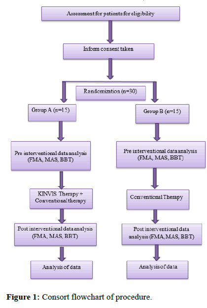
• The participants were allocated randomly into group A (experimental group) and group B (control group) (Figure 1).
Intervention
Group A: (Experimental groups: KINVIS+Conventional therapy): The participants in group A received kinesthetic illusion induced by visual stimulation (KINVIS) along with conventional therapy.
Study participants were familiarized individually with the KINVIS after randomization and prior to the intervention, according to the group to which they were assigned. To begin, the participants were briefed on the concepts of KINVIS and its application in lay term.
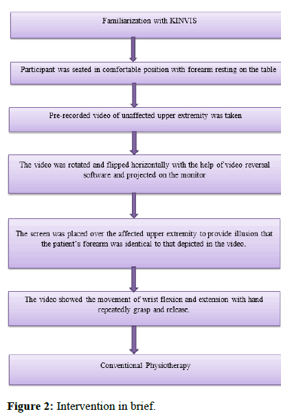
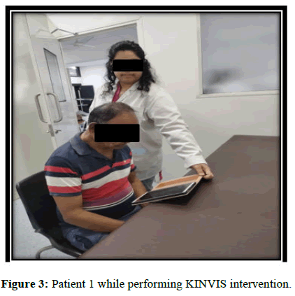
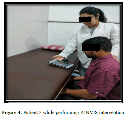
Patients were seated on a chair in a comfortable position with their forearm resting on the top of the table. A video of unaffected hand was recorded. Then a pre-recorded video of unaffected upper hand was flipped and rotated horizontally with the help of video reversal software and showed on a monitor. The video screen was placed over the affected forearm to provide the illusion that the patient’s forearm is identical to that depicted in the video. The video showed the movement of wrist flexion and extension with hand repeatedly grasp and release.
KINVIS was given for 20 minutes in two sets of 10 minutes with one-minute rest interval between the sets, five times a week frequency, for four weeks’ duration. This was followed by conventional physiotherapy (Figures 2-4).
Group B: (Conventional group): The goal of the conventional therapy group was to restore typical movement patterns and reduce spasticity.
Conventional therapy included:
• Static and dynamic control of positions.
• Balance exercises.
• Weight bearing exercises and weight shifts.
• ROM and stretching exercises.
• Activities of daily living training.
• Proprioceptive neuromuscular facilitation techniques.
• Neurodevelopmental facilitation techniques.
Conventional therapy was given for 45 minutes a day, 5-times-a-week frequency, for four week's duration.
Outcome measures
Fugl Meyer assessment scale-upper extremity: The FMAUE examines reflex activity, voluntary movements within, partially out and independent of synergies. The scale includes 33 items divided into 4 subscales: Shoulder/elbow (A, 18 items), wrist (B, 5 items), hand (C, 7 items) and coordination/speed (D, 3 items). Each item is scored on an ordinal 3-point scale, where 2 points are assigned when the movement is performed fully, 1 point when performed partially, and 0 points when the movement cannot be performed. A total score of 66 indicates better sensorimotor function. Both the intra and inter-rater reliability of the FMA-UE, by means of the Intraclass Correlation Coefficient (ICC), have demonstrated to be excellent, with reported values above 0.90.
Modified Ashworth scale: The modified Ashworth scale is a clinical scale used to assess muscle spasticity. The modified Ashworth scale is 5-point ordinal scale with 0 as no increase in muscle tone and 4 is considered as rigid. Tone assessment of wrist flexor muscles was done at the baseline and after 4 weeks of intervention.
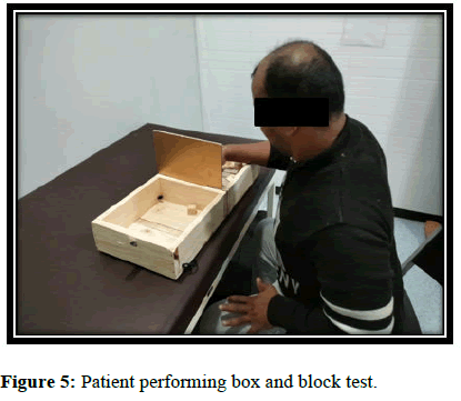
Box and block test: The Box and Block Test (BBT) is an outcome measure used to assess and monitor unilateral upper extremity manual dexterity. Box and Block set-up includes: A wooden box with dimensions 21.5 inches by 10.1 inches divided into two equal compartments by a wooden partition with a height of 6.0 inches. The box should be opened and placed on a table top in a horizontal orientation in front of the patient, who is seated in a chair. All cubes of 1 inches are placed on one side of the partition and the patient is asked to transfer as many blocks as they can in 60 seconds to the other side of the box. The blocks must be transferred one at a time, the patient must only use one hand and the hand should cross the partition in the transfer. The score is indicated by the number of blocks that can be transferred in 60 seconds.
Excellent test-retest reliability was found in the populations with, stroke (ICC=0.85 respectively). (Figure 5).
Statistical analysis
Statistical analysis of data was done using SPSS 27.0 version software. The results were considered significant at p<0.05 and Confidence Interval (CI) at 95%. Analysis was done using Kolmogorov Smirnov test, descriptive and inferential statistics using chi square test, student’s paired and unpaired t test. The Kolmogorov Smirnov test was used to assess the normality of data. For quantitative variables the mean and standard deviation was calculated and for categorical variables proportions were calculated. The data was represented using tables and in the form of visual impressions such as bar diagram.
Results
Total 30 participants based on the eligibility criteria were recruited in the study into 2 groups. Group A: Experimental group and group B: Control group.
The data was analyzed by taking 15 subjects in experimental group (group A) and 15 in control group (group B).
The results are based on all 30 participants. There were no dropouts during the study. The baseline characteristics of the participants are presented in Tables 1-4.
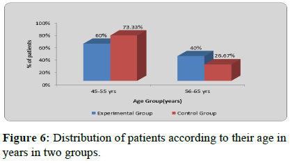
| Age group (yrs) | Experimental group | Control group | ê?2-value |
|---|---|---|---|
| 45-55 yrs | 9 (60%) | 11 (73.33%) | 0.6, P=0.43, NS |
| 56-65 yrs | 6 (40%) | 4 (26.67%) | |
| Total | 15 (100%) | 15 (100%) | |
| Mean ± SD | 53.20 ± 6.38 | 53.33 ± 5.40 | |
| Range | 45-64 yrs | 46-63 yrs |
Table 1: Distribution of patients according to their age in years in two groups.
| Gender | Experimental group | Control group | ê?2-value |
|---|---|---|---|
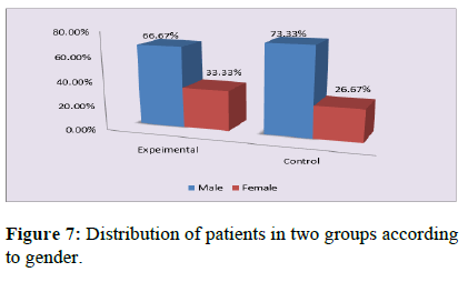
| Male | 10 (66.67%) | 11 (73.33%) | 0.15, P=0.69, NS |
| Female | 5 (33.33%) | 4 (26.6%) | |
| Total | 15 (100%) | 15 (100%) |
Table 2: Distribution of patients according to their gender in two groups.
| Type of stroke | Experimental group | Control group | ê?2-value |
|---|---|---|---|
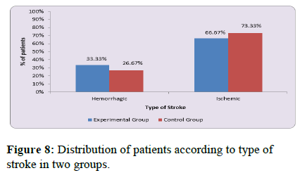
| Hemorrhagic | 5 (33.33%) | 4 (26.67%) | 0.15, P=0.69, NS |
| Ischemic | 10 (66.67%) | 11 (73.33%) | |
| Total | 15 (100%) | 15 (100%) |
Table 3: Distribution of patients according to type of stroke in two groups.
| Side affected | Experimental group | Control group | ê?2-value |
|---|---|---|---|
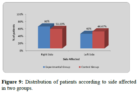
| Right side | 9 (60%) | 8 (53.33%) | 0.13, P=0.71, NS |
| Left side | 6 (40%) | 7 (46.67%) | |
| Total | 15 (100%) | 15 (100%) |
Table 4: Distribution of patients according to side affected in two groups.
Table 1 and Figure 6 represents the distribution of age of group A and group B which shows non-significant difference between the groups (‘p’=0.43).
Table 2 and Figure 7 depicts the gender distribution in group A and group B. In group A, total no. of participants in the study were 15 (male=10, female=5). In group B, total no. of participants in the study were 15 (male=11, female=4). The between group value shows no significant difference (p=0.69).
Table 3 and Figure 8 represents the distribution according to type of stroke of group A and group B which shown nonsignificant difference between groups (p value=0.69).
Table 4 and Figure 9 shows the distribution of affected side in stroke of group A and group B which showed non-significant difference between groups (p=0.71).
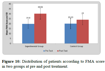
Mean FMA score at pre-treatment in experimental group was 19.80 ± 4.42 and at post-treatment it was 30.06 ± 5.39. By using student’s paired t test statistically significant difference was found in FMA score at pre and post treatment in experimental group (t=21.71, p=0.0001).
Mean FMA score at pre-treatment in control group was 20.13 ± 5.43 and at post-treatment it was 24 ± 5.29. By using student’s paired t test statistically significant difference was found in FMA score at pre and post treatment in control group (t=14.12, p=0.0001).
On comparing mean difference in FMA among patients of two groups by using student’s unpaired t test statistically significant difference was found in between two groups (t=11.7, p=0.001) (Table 5 and Figure 10).
| Group | Pre-treatment | Post-treatment | Mean difference | Student’s paired t-test t-value |
|---|---|---|---|---|
| Experimental group | 19.80 ± 4.42 | 30.06 ± 5.39 | 10.26 ± 1.83 | 21.71 P=0.0001, S |
| Control group | 20.13 ± 5.43 | 24 ± 5.29 | 3.86 ± 1.06 | 14.12 P=0.0001, S |
| Comparison of mean difference in two groups (Student’ unpaired t test) → | t-value | p-value | ||
| 11.71 | 0.001 | |||
Table 5: Distribution of patients according to FMA score in two groups at pre and post-treatment.
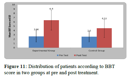
Mean BBT score at pre-treatment in experimental group was 2.66 ± 1.34 and at post-treatment it was 6.40 ± 2.32. By using student’s paired t test statistically significant difference was found in BBT score at pre and post treatment in experimental group (t=10.05, p=0.0001).
Mean BBT score at pre-treatment in control group was 2.60 ± 1.24 and at post-treatment it was 4.53 ± 2.13. By using student’s paired t test statistically significant difference was found in BBT score at pre and post treatment in control group (t=6.43, p=0.0001).
On comparing mean difference in BBT among patients of two groups by using student’s unpaired t test statistically significant difference was found in between two groups (t=3.77, p=0.001) (Table 6 and Figure 11).
| Group | Pre test | Post test | Mean difference | Student’s paired t-test t-value |
|---|---|---|---|---|
| Experimental group | 2.66 ± 1.34 | 6.40 ± 2.32 | 3.73 ± 1.43 | 10.05 P=0.0001, S |
| Control group | 2.60 ± 1.24 | 4.53 ± 2.13 | 1.93 ± 1.16 | 6.43 P=0.0001, S |
| Comparison of mean difference in two groups (Student’ unpaired t test) → | t-value | p-value | ||
| 3.77 | 0.001 | |||
Table 6: Distribution of patients according to BBT score in two groups at pre and post-treatment.
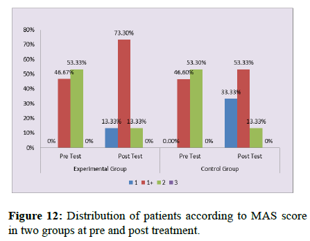
Modified Ashworth Scale score (MAS) 1, 1+ and 2 at baseline for experimental group was 0%, 46.6% and 53.3%respectively. MAS score 1, 1+ and 2 after 4 weeks of intervention was 13.3%, 73.3% and 13.3% respectively.
Modified Ashworth scale score 1, 1+ and 2 at baseline for control group was 46.6%, 13.3% and 53.3% respectively. MAS score 1.1+ and 2 after 4 weeks of intervention were 33.3%, 53.3% and 13.3% respectively (Table 7 and Figure 12).
| MAS score | Experimental group Pre test |
Experimental group Post test |
Control group Pre test |
Control group Post test |
ê?2-value |
|---|---|---|---|---|---|
| 1 | 0 (0%) | 2 (13.33%) | 7 (46.6%) | 5 (33.33%) | 18.07 P=0.006, S |
| 1+ | 7 (46.67%) | 11 (73.3%) | 2 (13.33%) | 8 (53.33%) | |
| 2 | 8 (53.33%) | 2 (13.33%) | 8 (53.33%) | 2 (13.33%) | |
| 3 | 0 (0%) | 0 (0%) | 0 (0%) | 0 (0%) | |
| Total | 15 (100%) | 15 (100%) | 15 (100%) | 15 (100%) |
Table 7: Distribution of patients according to MAS score in two groups at pre and post-treatment.
Discussion
The purpose of this pilot randomized control trial was to evaluate the effect of Kinesthetic Illusion Induced by Visual Stimulation (KINVIS) as an adjunct to conventional physiotherapy on hand function compared to conventional therapy alone in patients with chronic stroke. The outcomes measure used to evaluate hand function of the subjects were Fugl Meyer Assessment for Upper Extremity (FMA-UE), Box and Block Test (BBT) and Modified Ashworth Scale (MAS) for assessing tone.
The results of the study showed significant improvement in both experimental group and control group for FMA and BBT after 4 weeks of intervention. Between group analyses for FMA and BBT was significant with experimental group showing more statistically significant improvement as compared to the conventional group. The KINVIS group demonstrated notable improvement in wrist flexor muscle tone according to the Modified Ashworth Scale (MAS). Thus interpretating that kinesthetic illusion induced by visual stimulation along with conventional physiotherapy has added effects in improving hand functions in patients with chronic stroke
Ryosuke Takahashi et al. in his study investigate impact of repetitive KINVIS along with therapeutic exercise on hand function in patients with chronic stroke and found that the resistance to passive movement i.e. tones of wrist flexor muscle had reduced, and the motor function of the upper limb, including the finger, had improved following the intervention. In the study, total score of FMA and ARAT exhibited significant enhancement, with a median rise of 2.5 points. The results were similar to our study where kinesthetic Illusion Induced by Visual Stimulation (KINVIS) also showed significant benefits for improving hand function in experimental group i.e. improvement in FMA-UE and BBT score in chronic stroke patients. This might be due better formation of motor understanding and memory through watching a video of own body part. KINVIS is improved to have a psychological impact on recollection of movement in such patients and the kinesthetic feeling generated during KINVIS may improve the ability to produce voluntary motor output. Additionally, this process promotes the increased excitability of the mirror neuron system and areas of the motor network to improve motor learning in patients with stroke.
As previously mentioned, the results of our study showed improvement in Modified Ashworth Scale (MAS) for wrist flexor muscle tone in patients with chronic stroke after application of KINVIS. A similar effect was seen in the research study conducted by, Junpei Tanabe et al., on the paralyzed side ankle joint and composite spasticity score of ankle planter flexor muscles. There have been studies which indicate that KINVIS encourages the reciprocal engagement of muscles acting as agonist and antagonists. The study stated that the results of ankle dorsiflexion test and composite spasticity score was due to activation of the tibialis anterior and suppression of the triceps surae by KINVIS which may have improved the range of motion and smoothness of active ankle dorsiflexion. These findings are also supported by the results found in our study. The same mechanism may have shown better improvement in wrist flexor muscle tone in our study.
Fuminari Kaneko, et al., showed that KINVIS affects corticospinal tract excitability and induces motor control-associated cerebral network activation. The stimulation of frontoparietal neural network is linked to visual-motor tasks and the mirror neuron system, which is engaged in activation of motor association areas (pre-motor cortex, supplementary motor cortex) while performing KINVIS. Buccino, et al., also added that mirror neuron system in the human brain is connected to the process of learning through imitation or comprehending the significance of the movement. The Mirror Neuron System (MNS) plays a role in the process where action observation results in involving mechanisms such as visual observation, motor imagery, replication and learning. This procedure triggers neural plasticity by activating the brain MNS following stroke. Mirror neurons system activation has been found to contribute to improve motor functions in stroke patients these might be the reason that our study has shown significant improvement in experimental group. Hence, utilizing MNS-based training could potentially serve as a valuable novel approach for functional recovery post stroke.
Stroke results in abnormal inter-hemispheric interactions. Inter-hemispheric abnormalities develop after stroke as there is an over-activation or over use of the intact upper extremity. This hemispheric imbalance can be particularly improved by increasing the excitability of the affected hemisphere. Moreover, as postulated by Auriat, et al. improvement in interhemispheric interactions is assumed to be involved in recovery from paralysis after a stroke. KINVIS may, therefore, enhance recovery as it can selectively cause cortical excitability of the opposite hemisphere by inducing an illusion. KINVIS could therefore potentially trigger kinesthetic motor imagery, even among long-term chronic patients who have faced challenges in extending their hand for extended period of time. Katsuya Sakai, et al. in 2019 evaluated the effect of KINVIS on dorsiflexior dysfunction in stroke patient, found significant improvement in 10 MWTS and voluntary ankle dorsiflexion simultaneous to KINVIS as it leads to improvement in ability to adjust muscle output due to balance between the visual information obtained from the video, and somatosensory feedback information.
Observation of action or movement video on the screen with kinesthetic illusion could facilitate motor cortex excitability via intra-cortical excitatory circuits, indicating their relevance in motor learning. Prior researches conducted by Nojima, et al., demonstrated the relevance of visual feedback in the process of motor learning. Furthermore, a preceding investigation revealed that the vividness of the kinesthetic motor imagery saw a notable enhancement following illusion condition, in contrast to the observation condition. The functional connectivity during kinesthetic motor imagery was remarkably increased in the right frontal-parietal network (the right pre-motor cortex and right parietal lobe) after the illusion condition compared with that after the observation conditions.
The benefits of KINVIS is that unlike pure action observation, KINVIS includes the common neural activity of the planning stage of explicit motor imagery, without combining this with intentionally generated motor imagery. Significantly, KINVIS is carried out through embodied visual stimulation and holds clinical significance, as demonstrated in our study.
Conclusion
4 weeks of kinesthetic illusion induced by visual stimulation is found to be effective to improve hand function in patients with chronic stroke. However, on within group analysis, both the intervention group showed improvement in all parameters at the end of 4 weeks.
Strength of the Study
• This approach helped to minimize the workload and thereby increase their active participation in rehabilitation.
• Another positive point was the adherence to the treatment throughout, which is evident from all 30 patients completing the study without any dropouts
• This approach is easy to incorporate in neurological patients as it involves single task and easy to concentrate on a monitor screen.
Limitations
• This study only included chronic stroke patients and effectiveness of KINVIS was not evaluated in other type of stroke subjects.
• This study has not taken the long term follow-up of the patients so no conclusion can be drawn about the long term effects of KINVIS.
Future Scope
• A study with larger sample size with longer follow-up would be useful.
• Effect of KINVIS on different stages of stroke can be evaluated.
• A randomized clinical trial to compare the effects of KINVIS and mirror therapy on different neurological population can be evaluated.
• Effectiveness of KINVIS along with other combination techniques can be evaluated in neurological patients.
Clinical Implication
• Kinesthetic illusion induced by visual stimulation can be used as an adjunct along with conventional physiotherapy in Neurorehabilitation settings for improvement in hand functions in patients with chronic stroke.
• Use of this treatment with the other conventional training will substantially increase the active participation of patients with chronic stroke.
References
- Ojha PT, Basak S, Aglave V, Yadav J. Incidence of stroke in adults according to age, sex and subtypes in urban Indian population. Neurol Neurosci Rep. 2020;3:1-4.
- Yavuzer G, Selles R, Sezer N, Sütbeyaz S, Bussmann JB, et al. Selections From This Month in the Archives. Arch Physical Med Rehabil. 2008;89:1-17.
- Lim KB, Lee HJ, Yoo J, Yun HJ, Hwang HJ. Efficacy of mirror therapy containing functional tasks in poststroke patients. Ann Rehabil Med. 2016;40:629-636.
[Crossref] [Google Scholar] [PubMed]
- Fletcher-Smith JC, Walker DM, Sprigg N, James M, Walker MF, et al. ESCAPS study protocol: A feasibility randomised controlled trial of ‘early electrical stimulation to the wrist extensors and wrist flexors to prevent the post-stroke complications of pain and contractures in the paretic arm’. BMJ Open. 2016;6:e010079.
[Crossref] [Google Scholar] [PubMed]
- Hs J. Outcome and time course of recovery in stroke. Part II: Time course of recovery. The Copenhagen Stroke Study. Arch Phys Med Rehabil. 1995;76:406-412.
- Duncan PW, Goldstein LB, Matchar D, Divine GW, Feussner J. Measurement of motor recovery after stroke. Outcome assessment and sample size requirements. Stroke. 1992;23:1084-1089.
[Crossref] [Google Scholar] [PubMed]
- Parker VM, Wade DT, Hewer RL. Loss of arm function after stroke: Measurement, frequency, and recovery. Int Rehabil Med. 1986;8:69-73.
[Crossref] [Google Scholar] [PubMed]
- Wade DT, Langton-Hewer R, Wood VA, Skilbeck CE, Ismail HM. The hemiplegic arm after stroke: Measurement and recovery. J Neurol Neurosurg Psychiatry. 1983;46:521-524.
[Crossref] [Google Scholar] [PubMed]
- Roby-Brami A, Jarrasse N, Parry R. Impairment and compensation in dexterous upper-limb function after stroke. From the direct consequences of pyramidal tract lesions to behavioral involvement of both upper-limbs in daily activities. Front Hum Neurosci. 2021;15:662006.
[Crossref] [Google Scholar] [PubMed]
- Kamper DG, Fischer HC, Conrad MO, Towles JD, Rymer WZ, et al. Finger-thumb coupling contributes to exaggerated thumb flexion in stroke survivors. J Neurophysiol. 2014;111:2665-2674.
[Crossref] [Google Scholar] [PubMed]
- McPherson JG, Chen A, Ellis MD, Yao J, Heckman CJ, et al. Progressive recruitment of contralesional corticoâ?reticulospinal pathways drives motor impairment post stroke. J Physiol. 2018;596:1211-1225.
[Crossref] [Google Scholar] [PubMed]
- Kwah LK, Herbert RD. Prediction of walking and arm recovery after stroke: A critical review. Brain Sci. 2016;6:53.
[Crossref] [Google Scholar] [PubMed]
- Decety J, Grezes J. Neural mechanisms subserving the perception of human actions. Trends Cogn Sci. 1999;3:172-178.
[Crossref] [Google Scholar] [PubMed]
- Hanakawa T. Organizing motor imageries. Neurosci Res. 2016;104:56-63.
[Crossref] [Google Scholar] [PubMed]
- Kaneko F, Yasojima T, Kizuka T. Kinesthetic illusory feeling induced by a finger movement movie effects on corticomotor excitability. Neuroscience. 2007;149:976-984.
[Crossref] [Google Scholar] [PubMed]
- Okawada M, Kaneko F, Shindo K, Yoneta M, Sakai K, et al. Kinesthetic illusion induced by visual stimulation influences sensorimotor event-related desynchronization in stroke patients with severe upper-limb paralysis: A pilot study. Restor Neurol Neurosci. 2020;38:455-465.
[Crossref] [Google Scholar] [PubMed]
- Takahashi R, Koiwa M, Wataru ID, Okawada M, Akaboshi K, et al. visually induced kinaesthetic illusion combined with therapeutic exercise for patients with chronic stroke: A pilot study. J Rehabil Med. 2022;44:1-6.
[Crossref] [Google Scholar] [PubMed]



 The Annals of Medical and Health Sciences Research is a monthly multidisciplinary medical journal.