Gender Differences in Radiographic Analysis of the Mental Foramen among Urhobo People of Southern Nigeria
Department of Human Anatomy and Cell Biology, Western Delta State University Teaching Hospital, Cleveland, United States
2 Department of Radiology, Western Delta State University Teaching Hospital, Cleveland, United States
Received: 06-Oct-2022, Manuscript No. AMHSR-22-76644; Editor assigned: 10-Oct-2022, Pre QC No. AMHSR-22-76644 (PQ); Reviewed: 24-Oct-2022 QC No. AMHSR-22-76644; Revised: 22-Feb-2023, Manuscript No. AMHSR-22-76644 (R); Published: 01-Mar-2023, DOI: 10.54608.annalsmedical.2023.90
Citation: Godswill OO, et al. Gender Differences in Radiographic Analysis of the Mental Foramen among Urhobo People of Southern Nigeria. Ann Med Health Sci Res. 2023;13:479-481
This open-access article is distributed under the terms of the Creative Commons Attribution Non-Commercial License (CC BY-NC) (http://creativecommons.org/licenses/by-nc/4.0/), which permits reuse, distribution and reproduction of the article, provided that the original work is properly cited and the reuse is restricted to noncommercial purposes. For commercial reuse, contact reprints@pulsus.com
Abstract
Objective: Identi ication is foremost to forensic investigators and this includes determining race, age and sex of individuals. This investigation is pointed toward deciding sex from detailed observation of the Mental Foramen (MF) by comparing the Upper Limit (UF-L) and Lower Limit of the foramen (LF-L) to the lower limit of mandible.
Methods: This retrospective study investigated a hundred and ten clear radiographs without any pathology of 55 males and 55 females, between 20-60 years.
Results: Statistically signi icant differences (p<0.05) were obtained in all measurements between gender with the obtained Mean and SD values of 14.63 ± 2.59 in males UF-L and 13.60 ± 2.57 in female UF-L, also 11.78 ± 2.41 and 10.58 ± 2.31 in males and females LF-L respectively. Signi icantly higher values were noticed in males, and this establishes differences between genders.
Conclusion: Conclusively, the exact position of MF in reference to mandible is an additional radiographic method to determine gender. Result obtained will be of relevance to anatomist, forensic anthropologists and maxillofacial surgeons.
Keywords
Forensic anthropology; Mental foramen; Radiograph; Sex determination
Introduction
A cardinal focus of identification is gender determination. This is often necessary in medico legal situations where a person’s identity is in doubt and needs to be established. In the event of wars, accidents, incessant killings by bandits and discovery of mutilated body parts, identification is usually inevitable either for burial purposes or legal reasons. Gender determination and identification of individual is believed to be possible through different body parts such as long bones, pelvic bone and skull [1-3]. However the skull is termed the most accurate. Although, Identification of unknown bodies is always a challenge to forensic anthropologist, analysis of bound landmarks on bones may be a common approach employed by anthropologists and forensic scientist to resolve this puzzle and establish the gender and identity of unidentified persons. Several landmarks on the orbital bone, frontal bone, tooth and jaws in human skull and the pelvis are useful to forensic scientists and anthropologist indetermination of age, sex, and personal identification. Additionally, it has been established that the MF can be utilized in identification, particularly when only the skull is available for examination [4]. MF is found on the mandible and may be useful in determining gender. Mandible is assumed to be the strongest human bone and persists well in an exceedingly preserved state longer than the other bone as such could be useful for identification and gender determination when necessary [5]. The capacity to view the whole mandible permits a precise distinguishing proof and scrutiny of the foramen on radiographs. Al Jasser described MF as a space or an opening present on the anterior portion of the mandible. Usually, present on either side for passage of mental vein, artery, and nerves. Multiple MF may also occur, sometimes these foramen may be completely absent [6]. When radiographs obviously showing the foramen is taken, the positioning could be helpful in determining age, sex and ethnicity [7]. Regarding positioning and length, MF varies not only according to age, sex and ethnicity but also within the same race in different geographic regions and within the inhabitant of the similar geographical area [8]. Significance differences have shown that this foramen differs among individuals and thus useful in gender prediction of an unknown person. Hence studying MF divisibility in different ethnicity and its potential usefulness in sex prediction is worthy of note [9]. This study intend to assess the differences that exists between the MF of males and females and its usefulness in predicting gender among the Urhobo people of Southern Nigeria.
Materials and Methods
The study was retrospective in design, following ethical approval from the research and ethics committee of human anatomy department and ethical approval from university teaching hospital, Oghara, Delta state. One hundred and forty five radiographs were available, out of which 110 radiographs (55 radiographs for males and 55 for female) showing the foramen met the criteria and were adapted.
Inclusion criteria
• Available radiographs indicating age above 20 years.
• High quality radiograph.
Exclusion criteria
• Radiographs with malignancies.
• Unidentifiable foramen.
Method of data collection
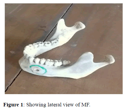
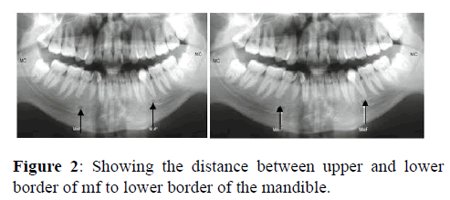
Measurement was achieved using computed radiographic films showing the foramen clearly. Quantitative data were collected from the mandible showing the foramen as it was measured (left side only). Lines were drawn through the upper and lower limits of the foramen and opposite line drawn from the tangents to the lower limit of the mandible utilizing Photoshop. Data was analyzed using unpaired t-test. Comparison was achieved on both sexes as P value was fixed at 0.05. P-values less than 0.05 were considered statistically significant (Figures 1 and 2).
Results
Table 1 Revealed significant gender difference. Males showed higher mean ± standard deviation in the distance measured from the upper border of the MF to the lower border of the mandible.
| Gender | Mean ± SD (mm) | Standard Mean Error (mm) | T-test value | P-value |
|---|---|---|---|---|
| Male | 14.63 ± 2.59 | 0.35 | 2.1 | 0.04 |
| Female | 13.60 ± 2.57 | 0.35 |
Table 1: Distance between upper limit of the MF to lower limit of the mandible (UF-L) between genders.
Table 2 showed significant gender difference. Males showed higher mean ± standard deviation in the distance measured from the lower border of the MF to the lower border of the mandible.
| Gender | Mean ± SD (mm) | Standard Mean Error (mm) | T-test value | P-value |
|---|---|---|---|---|
| Male | 11.78 ± 2.41 | 0.32 | 2.69 | 0.01 |
| Female | 10.58 ± 2.31 | 0.31 |
Table 2: Distance between lower limit of the MF to lower limit of the mandible (LF-L) between genders.
Discussion
Radiographic assessment of MF is useful in predicting gender as it plays an extremely useful role in identification [10]. Additionally, the MF has been reported to differ in location in diverse ethnic groups as well as gender wise [11]. From this study, UF-L were higher in males than in females. The finding is in harmony with the work of Al-Khateeb et al., who demonstrated critical contrasts in position and distance of MF in males and females, with males having higher measurements. Another study by Kusum and Sapna also supported the findings observed in this present study and expressed thatin all their linear measurements, males had higher measurements than females. A similar trend was also observed by Yosue et al., and Cagri et al., who showed that there are significant differences in location of MF between genders [12,13]. The observed high values in males may be attributed with the fact that in the adult phase, the rate and speed of bone growth are higher in men, so craniofacial dimensions in this gender are from 5% to 9% bigger relative to those of women. Longer length in males may be attributed to the fact that males enter puberty at a later year than females [14-17].
Conclusion
Also, our study reveals a significant variation in the inferior MF to lower border of the mandible (LF-L), hence shows higher values in males relative to females. The finding from this current study agrees with prior studies who reported that values of UF-Land LF-L were significantly high up in males. On the whole, the study showed definite sexual contrasts in dimensions of MF. Longer lengths of parameters measured often indicate males.
Disclosures/Conflicts of Interests
Nil
References
- KhanguraRK, Sircar K, Singh S, Rastogi V (2011) Sex determination using mesiodistal dimension of permanentmaxillary incisors and canines. J Forensic Dent Sci. 2011;3:81-85. [Crossref] [Google Scholar] [PubMed]
- UthmanAT, Al-Rawi NH, Al-Timimi JF. Evaluation of foramen magnum in gender determination usinghelical CT scanning. DentomaxillofacRadiol. 2012; 41:197-202. [Crossref] [Google Scholar] [PubMed]
- EbeyeAO, Okoro GO, Ikubor EJ. Radiological assessment of age from epiphyseal fusion atthe wrist and ankle in Southern Nigeria. Forensic Sci Int. 3:1-11. [Google Scholar]
- MamerhiET, Godswill OO. Anthropometric study of the frontal sinus on plainradiographs in Delta State University Teaching Hospital. J Exp Clin Med. 2018; 17:49:52. [Google Scholar]
- EnaohwoTM, Okoro OG. Morphometric study of hypoglossal canal of occipital bonein dry skulls of two states in southern Nigeria. Bangladesh J Med Sci. 19:670-672. [Google Scholar]
- Sahni P,Patel RJ, Shylaja, HM, Anil P. Gender determination using panomograhic (OPG) Analysis ofmental foramen in North Gujarat population. Med Res Chron. 2015; 23:943-971. [Google Scholar]
- Kusum S,Sapna S. Gender determination by mental foramen using linearmeasurements on radiographs: a study in Haryana population. Ind J Forensic Med Toxicol. 2016; 10:45-49. [Google Scholar]
- ChandraA, Singh A, Bandy M, Jaiswal R, Agnihotri A. Determination of sex by radiographic analysis of mentalforamen in North Indian population. J Forensic Dent Sci. 2013;5:52-55. [Crossref] [Google Scholar] [PubMed]
- AlJasser NM, Nwoku AL. Radiographic study of the mental foramen in a selectedSaudi population. DentomaxillofacRadiol. 1998; 27:341-343. [Crossref] [Google Scholar] [PubMed]
- PhillipsJL, Weller RN, Kulild JC. The mental foramen: Size, orientation, and positionalrelationship to the mandibular second premolar. J Endod. 1990; 16:221-223. [Crossref] [Google Scholar] [PubMed]
- PokhrelP, Bhatnagar R. Position and number of mental foramen in dry humanmandibles: Comparison with respect to sides and sexes. OA Anatomy. 2013;1:1-6.
- SingalK, Sharma S. Gender determination by mental foramen using linearmeasurement on radiographs: a study in Haryana population. Indian J Forensic Med Toxicol. 2016; 5:68-73. [Google Scholar]
- SaxenaS, Sharma P, Gupta N. Experimental studies of forensic odontology to aid in theidentification process. JForensic Dent Sci. 2010;2:69-76. [Crossref] [Google Scholar] [PubMed]
- AlKhateeb T, Al HadiHamasha A, Ababneh KT. Position of themental foramen in a northern regionalJordanian population. SurgRadiol Anat. 2007;29:231-237. [Crossref] [Google Scholar] [PubMed]
- Yosue T,Brooks SL. The appearance of mental foramina on panoramic andperiapical radiographs. OralSurg, Oral Med, Oral Path. 1989;68:488-492. [Crossref] [Google Scholar] [PubMed]
- Cagri U,Cihan B, Ismail S, Ali MA, Yusuf ZA. Bone height measurement of maxillary and mandibular bonesin panoramic radiographs of edentulous patients. J Clin Exp Dent. 2011;3:5-9. [Google Scholar]
- Sura A.Jamali A. Sex determination using linear measurements related to themental and mandibular foramina vertical positions on digital panoramic images. Bangladesh J Dent Res Educ. 2011;23:1-6.[Google Scholar]


 The Annals of Medical and Health Sciences Research is a monthly multidisciplinary medical journal.