Heart and Lung Hydatid Cyst in a Pregnant Woman: A Case Report
Citation: Rostami A, et al. Heart and Lung Hydatid Cyst in a Pregnant Woman: A Case Report. Ann Med Health Sci Res. 2017; 7: 373-376
This open-access article is distributed under the terms of the Creative Commons Attribution Non-Commercial License (CC BY-NC) (http://creativecommons.org/licenses/by-nc/4.0/), which permits reuse, distribution and reproduction of the article, provided that the original work is properly cited and the reuse is restricted to noncommercial purposes. For commercial reuse, contact reprints@pulsus.com
Abstract
We reported a 23-year-old female, 2 months pregnant presented with cough and blood streaked sputum (Hemoptysis) since two weeks ago. Chest CT scan showed simultaneous multiple cystic lesions in the lungs and heart. After therapeutic abortion, the patient was operated via median sternotomy incision and all hydatid cysts were extracted of the lung and heart. The patient was discharged after one week with good condition. The abdominal and pelvic sonography of the patient was also normal with no cystic lesions. No endo-bronchial lesions or malignant cells were reported in bronchoscopy
Keywords
Echinococcus granulosus; Lung; Cyst; Heart
Introduction
Hydatid cyst is a common parasitic disease between humans and animals (zoonosis) considered as an endemic disease in many regions of the world including South America, Africa, Greece, Turkey, some areas around the Mediterranean Sea, the Middle East, India, China, Australia, New Zealand, and Iran. [1] This cyst is caused by a parasite called Echinococcus granulosus which matures in the intestine of dogs (main host) or other canines such as wolves, foxes, etc. The rate of hydatid cyst development in lungs is three times as much as hydatid cyst in liver. [2,3] Pulmonary hydatid cysts usually have no symptoms except for the cases when they grow really large and cause coughs or hemoptysis. The side effects of pulmonary hydatid cyst emerge only when it ruptures inside bronchial tree or Pleura. [1-4] The most common symptoms of cardiac hydatid cyst are dyspnea angina and heart beat caused by the pressure put on the coronary artery and cardiac conduction system by the cyst. [5]
The cyst may rupture inside bronchial tree, peritoneal and pericardial cavities. If this rupture takes place inside the abdomen, it may result in cyst diffusion or the potentially lethal reaction of Anaphylactic. [4,5] Following the liver, lung is the second most common site of hydatid cyst in humans. The clinical presentation of pulmonary hydatid cyst depends on its size and location. The first symptom of pulmonary hydatid cyst is nonproductive cough. Other symptoms include chest cage pain, short breath and hemoptysis. Mild hemoptysis is a relatively common clinical symptom which may be caused by bronchial erosions or bronchial pressure and obstruction with cyst infection. Central hydatid cysts usually have a higher chance of hemoptysis. [4,5] Pulmonary hydatid cyst infects only one lobe (lower lips) in 72% of the cases while the simultaneous infection of both the liver and lung has been reported in 5 to 13% of the cases. The bilateral prevalence of pulmonary hydatid cyst has been reported to be around 2 to 30 %. [5] Chest CT scan is particularly valuable to discover small cysts (1 to 2 cm), complicated cysts of the lung and Mediastinum. Serologic tests are wrongly negative in 40% of lung cysts and 50% of cardiac cysts. [5] Occurrence of hydatid cyst in the heart is a rare phenomenon (0.02 to 2%). Hydatid cyst during pregnancy is also very rare. [6]
Hydatid cyst may reside in cardiac or pericardial cavities and cause myocardial ischemia, disrupted heart conduct, outlet obstruction and heart failures by putting pressure on coronary arteries. If cardiac hydatid cyst is not treated properly, it may rupture inside the heart or pericardium and result in pulmonary embolism, pericardial or systematic tamponade. Thus, the immediate treatment of coronary hydatid cyst is a necessity. [7]
The clinical image of cardiac Echinococcus depends on the site and intensity of the disruption of surrounding structures. Cardiac hydatidosis may represent itself through a nonspecific face such as weight loss, fever, dizziness, lethargy and heartbeat. [4] It is possible for the patient with cardiac hydatid cyst to have no symptoms at all (10%). Following the hydatid cyst rupture of the right hand side of the heart, the progressive pulmonary hypertension takes place immediately. This is due to the embolization of a large number of Scolexes inside the pulmonary blood circulation. [8]
Trans-esophageal echocardiography (TEE) is an elective diagonal procedure for cardiovascular hydatid cyst. [9] Cardiac or pulmonary hydatid cyst is mostly treated through operation. This operation seeks to eradicate the parasite, prevent cyst rupture, prevent diffusion of its content during the operation, destroy cyst’s remaining cavity and maintain pulmonary parenchyma. [5-10] The operation includes Enucleation, Pericystectomy, Capitonnage, segmental resection, lobectomy and (rarely) pneumonectomy and standard incision in posterolateral thoracotomy of lung cyst. Of all protoscolicidal fluids used during hydatid cyst extraction, none is ideally useful and assuring. One of these useful solutions is 20% hypertonic normal saline. [11] The elective incision in operating the bilateral lung hydatid cyst or simultaneous heart and lung cyst is transsternal incision (clamshell incision) or median sternotomy. [5] Serological monitoring, echocardiography, chest radiography, and, if necessary, chest CT scan during the 5 years following the operation in patients with both heart and lung cysts are recommended to trace the patients. [12] The aim of case report was to evaluate of heart and lung hydatid cyst in a pregnant woman
Case Presentation
A 23-year old woman presented from the countryside to doctor 2 weeks after pregnancy complaining about chest pain and sever coughs with hemoptysis. The patient was also complaining about weight loss (7 kg) and nocturnal sweating during this period. The patient claims to have experienced similar symptoms 3 months ago, but an outpatient treatment improved his health. The initial examinations found the patient to be anemic with normal sounds in lungs. As for the hearing of heart, murmur II/VI is heard in heart apex and the left side of sternum. The examination of the abdomen and organs was normal. Written informed consent was obtained from the patient for publication of this case report and any accompanying images
Laboratory tests
Sputum smear of the patient in terms of acid-fast bacilli (AFB) conducted three times was negative.
Chest cage radiography
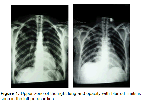
The picture of a density is observed in the upper zone of the right lung [Figure 1] . Opacity with blurred limits is seen in the left paracardiac which has not blurred the left side of the heart [Figure 1].
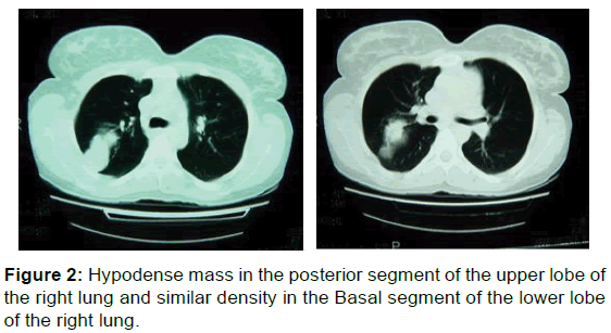
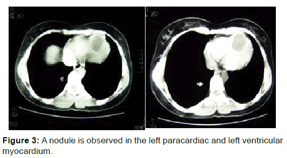
The image of a hypodense mass with a dimension of 2.5 × 3.2 cm in the posterior segment of the upper lobe of the right lung is observed [Figure 2] . A similar density with a size of 0.8 × 1 cm is seen in the Basal segment of the lower lobe of the right lung [Figure 2] . The image of a nodule with the size of 0.8 × 1.5 cm is observed in the left paracardiac, while an image of 2 other hypodense masses in pericardium and another hypodense mass with the size of 4 × 3 cm is observed in left ventricular myocardium [Figure 3] .
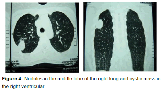
The results of chest HRCT reported a 3.5cm mass in the posterior segment of the upper lobe of the right lung and 25 and 12 mm nodules in the medial segment of the middle lobe of the right lung and the lingular lobe of the left lobe beside a 4 cm cystic mass in the right ventricular [Figure 4].
Echocardiography
A 37 × 36 mm hypoechoic lesion is observed in the apical septum of the heart with more tendencies to the right side which seems to be a mere number.
The abdominal and pelvic sonography of the patient was also normal with no cystic lesions.
No endo-bronchial lesions or malignant cells were reported in bronchoscopy and BAL.
Discussion
The patient, initially diagnosed with cold, underwent an outpatient treatment with symptoms of cough and vague chest cage pains. As other symptoms such as dyspnea and traces of blood were observed in her sputum, chest radiography was prescribed. As the patient was pregnant, preparing the radiography was delayed and this issue delayed the diagnosis and treatment of patient. In any patient suspicious of hydatid cyst, liver and lung examination in terms of affliction with this disease is of particular importance.
On the other hand, the patient has lost 7 kilograms over the last months following one week of cold-like symptoms. As various papers indicate, patients with heart hydatid cyst may also experience weight loss. [7,13] The other point is that observation of several opacities with flat sides in chest radiography suggest hydatid cyst for the patient confirmed by CT scan of patient’s chest. Several hydatid cysts of the heart and the upper and lower lobes of the lung are observed in chest radiography and CT scan. An important diagonal point in the patient is transformation of left side of the heart in the chest which may be used as interesting and guiding principles in simultaneous diagnosis of heart and lung hydatid cyst in the patient.
However, heart hydatid cyst is usually concealed due to the high density of the heart. In some cases, the site or large size of the cyst may alter the heart meter. Such cases particularly in endemic regions will result in diagonal suspicion. [14] The other point to be remembered is that the artifact caused by heart movement is a major setback of CT scan in assessing cardiac hydatid cyst. Perhaps one of the most important tools of complementary diagnosis of hydatid cyst in these patients is to use chest MRI instead of chest CT scan. In compliance with the radiologist’s recommendations, chest HRCT was also conducted despite the fact that the patient was pregnant. [15]
Only one 36 × 37 mm hypoechoic lesion in the apical septum of the right ventricular was mentioned in patient’s echocardiography. However, we observed at least 3 hydatid cyst in heart wall during the exploration. Echocardiography also studies the compressing effect of cyst on heart’s performance. It is possible that chest MRI may be more accurate for studying and determining the number of heart hydatid cysts. The difference between the number of heart hydatid cysts reported in echocardiography and the number of heart hydatid cysts during the operation may be attributed to the fact that no MRI was conducted for this patient. [6-16]
As for diagnosing hydatid cyst serology and keeping in mind the fact that both heart and lung hydatid cysts were healthy, the negative result of anti-Echinococcus antibody test can be attributed to cyst wall antibody’s failure to penetrate into patient’s serum and negation of the test. In more than 30% of the patients suffering from different types of parasites (including hydatid cyst), Eosinophilia has been reported to be more than 7% (this variable was reported 2% for this patient). [1,10] Considering patient’s pregnancy and the necessity of an immediate operation on heart hydatid cyst and the teratogenic effect of albendazole, therapeutic abortion was prescribed by gynecologist. [17]
As a result, the patient underwent an operation 2 weeks following the commencement of albendazole. To shorten the time of using heart and lung pumping device, lung hydatid cyst was removed first. Next, the patient was put on artificial lung and heart pumping device and 3 heart hydatid cysts were also enucleated. Hypertonic saline was used to prevent diffusion of hydatid cyst and reduce the possibility of relapsing during the operation.
Conclusion
In summary, as hydatid cyst may infect every organ, we should always be suspicious of hydatid cyst if we encounter patients with single or several cystic lesions in one or several organs, particularly in endemic regions. If a patient is diagnosed with hydatid cyst, it is necessary to examine other organs particularly liver and lung for affliction with hydatid cyst. Bronchoscopy is of no help in diagnosing lung hydatid cyst, but it is quite necessary in cases of productive cough hemoptysis or hydatid cyst rupture.
Conflict of Interest
All authors disclose that there was no conflict of interest.
REFERENCES
- Geller DA, Goss JA, Anderson DK. Pollock Schwartz’s principles of surgery Ninth edition New York MC Grew Hill 2010; p: 1116.
- Angelica MD, Fong Y. The liver sabiston textbook of surgery 18th edition. Philadelphia Saunders, 2008; 1494-1496.
- Abu-Eshy SA. Some rare presentations of hydatid cyst (Echinococcus granulosus), king Saud university college of medicine, Abba, Kingdom of Saudi Arabia JR. Coll Sury, 1998: 43: 347-352.
- Kojundzic S, Dolic K, Buca A. Hydatid Disease with ultiple Organ Involvement: A Case Report Macedonian Journal of Medical Sciences, 2010; 3: 154-158.
- Harlaftis NN, Aletras HA, Symbas PN. Hydatid disease of the lung. In: Shields TW (edr.) General Thoracic Surgery. Lippincott Williams and Wilkins, Philadelphia. 2005; 1298-1307.
- Polat P, Kantarci M, Alper F. Hydatid disease from head to toe. Radiographics 2003; 23:475-494
- Ozbar C, Ozsoyler I, Arkci MK. Cardiac Hydatid cyst – ozbek Department of cardiovascular surgery, Izmir Ataturk Education and research hospital, Asian Cardiovascular thoracic Ann 2000; 8: 375-377.
- Jannati M, Nematti MH, Zamani J. Asymptomatic cardiac Hydatid cyst. Cardiac surgery ward, Faghihi Hospital Shiraz University of Medical Sciences. IJMS 2006; 31: 23-27.
- Leila A, Laroussi L, Abdennadher M. A cardiac hydatid cyst underlying pulmonary embolism: A case report. Pan Afr Med J 2011; 8: 12.
- Ghaemi M, Soltani E. Bilateral pulmonary hydatidosis after traumatic rupture of lung hydatid cyst in a child: report of a case. The Internet Journal of Surgery 2008; 20: 1-5.
- Takriti A, Hossin J, Jourabian M. Hydatid cyst in the cardiac papillary muscle of the tricuspid valve. Iran J Radiol 2010; 7: S7.
- Dursun M, Terzibasioglu E, Yilmaz R. Cardiac hydatid diease:CT and MRI findings. AJR Am J Roentgenol 2008; 190: 226-232.
- Soleimani A, Sahebjam M, Marzban M, Shirani S, Abbasi A. Hydatid cyst of the right ventricle in early pregnancy. Echocardiography 1998; 25: 778-870.
- WHO Informal Working Group on Echinococcosis. Guidelines for treatment of cystic and alveolar echinococcosis in humans. Bull World Health Organ 1996; 74: 231-242.
- Can D, Oztekin O. Hepatic and splenic hydatid cyst during pregnancy: A case report. Arch Gynecol Obstet 2003; 268: 239-240.
- Rahman MS, Rahman J, Lysikiewicz A. Obstetric and gynaecological presentations of hydatid disease. Br J Obstet Gynaecol 1982; 89: 665-670.
- Bartoloni C, Tricerri A, Guide L. The efficacy of chemotherapy with mebendazole in human cystic echinococcosis: Long-term follow-up of 52 patients. Ann Trop Med Parasitol 1992; 86: 249-256.


 The Annals of Medical and Health Sciences Research is a monthly multidisciplinary medical journal.