Histopathological Review of Nephrectomy Specimens in Ile-Ife: A 10-Year Retrospective Review
2 Teaching Hospital's Complex, Obafemi Awolowo University, Ile Ife, Nigeria
Received: 02-Oct-2022, Manuscript No. AMHSR-22-76401; Editor assigned: 05-Oct-2022, Pre QC No. AMHSR-22-76401 (PQ); Reviewed: 19-Oct-2022 QC No. AMHSR-22-76401; Revised: 27-Jan-2023, Manuscript No. AMHSR-22-76401 (R); Published: 09-Feb-2023
Citation: Abiola AA, et al. Histopathological Review of Nephrectomy Specimens in Ile-Ife: A 10-Year Retrospective Review. Ann Med Health Sci Res. 2023;13:446-450
This open-access article is distributed under the terms of the Creative Commons Attribution Non-Commercial License (CC BY-NC) (http://creativecommons.org/licenses/by-nc/4.0/), which permits reuse, distribution and reproduction of the article, provided that the original work is properly cited and the reuse is restricted to noncommercial purposes. For commercial reuse, contact reprints@pulsus.com
Abstract
Aim and objectives: The study aims to determine the common histopathological findings in the nephrectomy specimens received at the O.A.U.T.H.C. Ile-Ife over 10 years period.
Method: This is a cross-sectional retrospective histopathologic review of nephrectomy specimens received at the department of morbid anatomy and forensic medicine, O.A.U.T.H.C Ile- Ife from May 2012 to May 2020.
Results: There were 72 nephrectomy specimens seen in our histopathology department over the period of the study, 41 (56.9%) were females and 31 (43.1%) were males with an F:M ratio of 1.3:1. Seventy-six percent of the cases were malignant while 23.6% were benign. Renal cell carcinoma was the commonest indication for performing nephrectomy in the study period accounting for 28 cases (38.9%) followed by Wilms tumor which constitutes 24 of the cases (33.3%) out of the total. Cystic diseases of the kidney accounted for most (70.6%) benign conditions seen in this study followed by angiomyolipoma (11.8%). Fifty percent of the nephrectomies were done on the left kidney while 48.6% were done on the right kidney and 1.4% were bilateral.
Conclusion: Neoplastic lesions were commoner than non-neoplastic lesions in the nephrectomy specimens received during the study period. Renal cell carcinoma was the most common neoplasm seen and cystic diseases of the kidney were the most common non-neoplastic lesion observed.
Keywords
Neoplastic lesions; Forensic medicine; Morbid anatomy; Histopathological indings; Nephrectomy specimens
Introduction
Kidneys are paired bean shaped organs that sub-serve several functions in the body ranging from excretory, endocrine and metabolic functions amongst others. Several pathologies can affect the kidneys whose treatments include either simple or radical nephrectomy [1]. Nephrectomy is a surgical procedure that is performed for total or partial removal of the kidneys. The indications range from infections, benign and malignant conditions of the kidney. The indications vary according to age groups and geographical locations depending on the prevalent urological diseases [2]. Renal cell carcinoma is the most common malignant indication for nephrectomy in adults, while nephroblastoma is the most common malignant indication in children. Non-neoplastic conditions such as chronic pyelonephritis remains the most common indication for performing nephrectomy in many hospitals particularly in less developed countries where many renal diseases that are amenable to treatment are not detected on time due to poor diagnostic facilities thereby resulting in irreversible damage. Cystic renal diseases which may be congenital or acquired as well as donor renal transplant procedures are common indications for nephrectomy in some environment. Data on the patterns of diseases, age and gender distribution, side and common histological findings in nephrectomy specimens in our environment remains scanty. Hence, we set out to determine these parameters in our hospital.
Materials and Methods
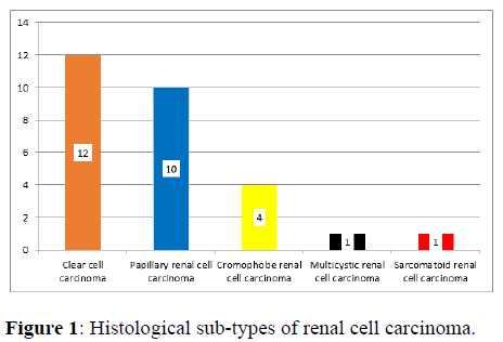
This was a retrospective cross-sectional study of all nephrectomy specimens received at the department of morbid anatomy and forensic medicine from May 2012 to May 2020. A total of 72 nephrectomy specimens were received in the department over a 10 years period. The histological request cards were retrieved and studied and relevant clinical information such as age, gender, histological diagnosis, laterality and location of the lesions were noted. The data obtained was analysed using SPSS version 16 (Figure 1).
Preparation for histology
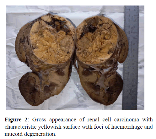
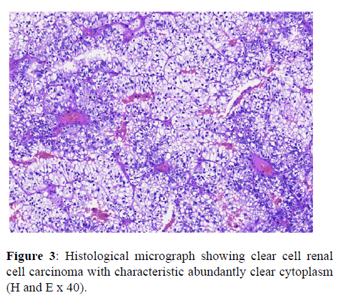
The tissue is then prepared for viewing under a microscope using either chemical fixation or frozen section. If a large sample is provided e.g. from a surgical procedure then a pathologist looks at the tissue sample and selects the part most likely to yield a useful and accurate diagnosis this part is removed for examination in a process commonly known as grossing or cut up. Larger samples are cut to correctly situate their anatomical structures in the cassette. Certain specimens (especially biopsies) can undergo agar pre-embedding to assure correct tissue orientation in cassette and then in the block and then on the diagnostic microscopy slide. This is then placed into a plastic cassette for most of the rest of the process (Figures 2 and 3).
Chemical fixation
In addition to formalin, other chemical fixatives have been used. But, with the advent of Immunohistochemistry (IHC) staining and diagnostic molecular pathology testing on these specimen samples, formalin has become the standard chemical fixative in human diagnostic histopathology. Fixation times for very small specimens are shorter, and standards exist in human diagnostic histopathology.
Procession
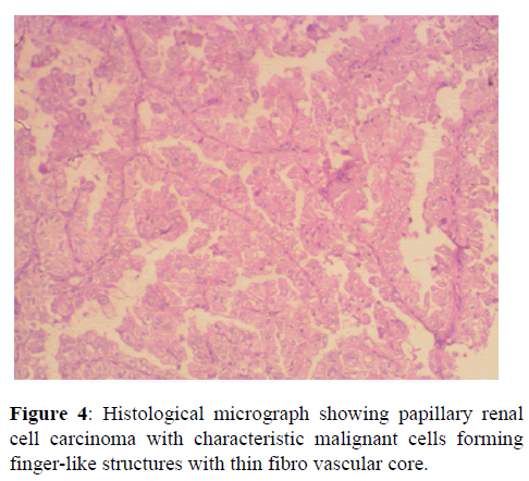
Water is removed from the sample in successive stages by the use of increasing concentrations of alcohol [3] xylene is used in the last dehydration phase instead of alcohol this is because the wax used in the next stage is soluble in xylene where it is not in alcohol allowing wax to permeate (infiltrate) the specimen. This process is generally automated and done overnight. The wax infiltrated specimen is then transferred to an individual specimen embedding (usually metal) container. Finally, molten wax is introduced around the specimen in the container and cooled to solidification so as to embed it in the wax block [4] this process is needed to provide a properly oriented sample sturdy enough for obtaining a thin microtome section(s) for the slide. Once the wax embedded block is finished, sections will be cut from it and usually placed to float on a water bath surface which spreads the section out. This is usually done by hand and is a skilled job (histotechnologist) with the lab personnel making choices about which parts of the specimen microtome wax ribbon to place on slides. A number of slides will usually be prepared from different levels throughout the block. After this the thin section mounted slide is stained and a protective cover slip is mounted on it. For common stains, an automatic process is normally used; but rarely used stains are often done by hand (Figure 4).
Results
A total of 72 nephrectomy cases were studied. 58 (80.6%) of these cases were neoplastic and 14 (13.4%) were non-neoplastic. There were 41 (56.9%) females and 31 (43.1%) males giving a female to male ratio of 1.3:1. The age range was from 4 months to 84 years and the modal age group was 0-10 years (43.1%). Out of the 72 nephrectomies done, 36 (50%) were done on the left kidney while 35 (48.6%) were done on the right kidney and 1 (1.4%) was bilateral. The histological diagnosis in 55 (76.4%) of the cases were malignant while 17 cases (23.6%) were benign. Twenty-four (43.6%) of the malignant cases were due to Wilms tumor while renal cell carcinoma accounted for 28 (50.9%) of the cases, other malignant tumors seen were 1 case (1.9%) each of Non-Hodgkin lymphoma, clear cell sarcoma and urothelial carcinoma of renal pelvis. Cystic diseases of the kidneys were the most common benign renal lesions observed accounting for 12 (70.6%) cases and they include; 6 cases of multi-cystic renal dysplasia, 4 cases of adult polycystic renal disease, 1 case each of autosomal polycystic renal disease and acquired renal cystic disease.
The other benign lesions observed include; 2 cases of angiomyolipoma, and 1 case each of mesoblastic nephroma, transplant rejection, and renal abscess respectively (Tables 1 and 2).
| Table 1: Histopathological findings in nephrectomy specimen. | ||
|---|---|---|
| Histological diagnosis | Frequency | Percentage |
| Polycystic kidney disease | 5 | 6.90% |
| Nephroblastoma | 24 | 33.30% |
| Renal cell carcinoma | 28 | 38.90% |
| Angiomyolipoma | 2 | 2.80% |
| Multi-cystic renal dysplasia | 6 | 8.30% |
| Mesoblastic nephroma | 1 | 1.40% |
| Non-Hodgkin lymphoma | 1 | 1.40% |
| Renal abscess | 1 | 1.40% |
| Transplant rejection | 1 | 1.40% |
| Clear cell sarcoma | 1 | 1.40% |
| Transitional carcinoma | 1 | 1.40% |
| Acquired cystic renal disease | 1 | 1.40% |
| Total | 72 | 100% |
| Table 2: Frequency of nephrectomy specimens across age groups. | ||
|---|---|---|
| Age groups (years) | Frequency | Percentage |
| 0-10 | 31 | 43.10% |
| 11-20 | 2 | 2.80% |
| 21-30 | 10 | 13.80% |
| 31-40 | 6 | 8.30% |
| 41-50 | 3 | 4.20% |
| 51-60 | 10 | 13.90% |
| 61-70 | 7 | 9.70% |
| 71-80 | 2 | 2.80% |
| 81-90 | 1 | 1.40% |
| Total | 72 | 100% |
Discussion
The histological findings in nephrectomy specimens tend to vary according to age and geographical location globally. The most common indication for performing nephrectomy in this study were neoplastic renal diseases accounting for 80.6% of the cases. This is in agreement with studies done by Onyeanunam et al. in South-south Nigeria and Be Island in Norway who reported that neoplastic renal diseases accounted for 57.9% and 75% of nephrectomy specimens. This is however in contrast with what Shaila and Bharti described in India where they reported non neoplastic conditions particularly chronic nonspecific pyelonephritis accounting for 77.6% and 88.6% of the indications for nephrectomy respectively [5,6]. In this study, 94.8% of the neoplastic cases were malignant while 5.2% were benign. However, Swarnlata et al reported that 58.45% and 41.53% of their nephrectomy surgeries were due to malignant and benign neoplastic conditions respectively [7]. Renal cell carcinoma is the most commonly reported malignant tumor in nephrectomy specimens in adults. The renal cell carcinoma constituted 43.6% of all malignant tumors in adults in this study. This is comparatively lower than 78.6% that was reported by Swarnlata, et al. Clear cell variant of renal cell carcinoma was the most common histological subtype of renal cell carcinoma in this study (42.9%) followed by papillary renal cell carcinoma (35.7%) and chromophore renal cell carcinoma (14.3%). This is slightly lower than what was previously reported in our center by Salako, et al. who documented that clear cell variant of renal cell carcinoma accounted for 60.8% of renal cell carcinoma [8]. The difference may be explained by the fact that they included some cases obtained at the autopsy to their cohort. It is however very similar to the finding by Badmus, et al. in the same center who reported clear cell variant to be responsible for 46.2% of all renal cell carcinoma 13 years ago [9]. The nephrectomy specimen received in this study were slightly more commonly done on the left than on the right while it is bilateral in a case and this is similar to finding from previous studies in our center. There were 41 (56.9%) females and 31 (43.1%) males giving a female to male ratio of 1.3:1. This is in contrast to the works by Badmus et al and El Malik, et al. who reported female to male ratio of 1:2 and 1:1.9 respectively [10]. This may be due to the fact that these studies only reviewed nephrectomy in adults as against our study where we reviewed nephrectomy in all age groups. Most of the nephrectomy cases were obtained from patients less than 10 years and most of these cases were done in patients with nephroblastoma followed by multi-cystic renal dysplasia. Nephroblastoma was the most common renal malignancy in children in this study and this is in concordance with the finding by Basir, et al. and Uchechukwu [11,12]. The upper pole was the commonest location of the renal tumor accounting for 40.3% of the cases while the entire kidney was involved in 25% of cases. This is similar to the studies by Basir and Popat, et al. [13].
Conclusion
In conclusion, neoplastic renal diseases were the most common indications for nephrectomy in our center. Renal cell carcinoma was the most common malignant tumor seen in the nephrectomy specimens in adult while Wilms tumor was the most common malignant tumor in the nephrectomy specimen in children. Unlike in many studies, there were more cases in women than in men.
Authors Contributions
• Adefidipe AA (Research design, data analysis and manuscript writing).
• Adelusola KA (Definition of intellectual content and Manuscript editing).
• Komolafe AO (Statistical analysis and manuscript editing).
• Adefehinti O (Manuscript review).
• Ogunrinde OV (Data collection).
• Olorunsola I.S (Data acquisition and analysis).
• Alade O.T (Manuscript review and literature search).
The manuscripts has been read by all the authors.
References
- Oh JJ, Byun SS, Lee SE, Hong SK, Lee ES. Partial Nephrectomy versus radical nephrectomy for non -metastatic pathological T3 a renal cell carcinoma:A multi-institutional comparative analysis. Int J Urol. 2014; 21:352-357. [Crossref] [Google Scholar] [PubMed]
- Datta B, Moitra T, Chaudhury DN, Halder B. Analysis of 88 nephrectomies in a Rural Tertiary Care Center of India. Saudi J Kidney Dis Transpl. 2012;23:409-413. [Google Scholar] [PubMed]
- Onyeanunam NE, Olatunde EA. Open nephrectomy: Experience in a Nigerian Teaching Hospital. JAMMR. 2017;24:1-8. [Google Scholar]
- Beisland C, Medby PC, Sander S, Beisland HO. Nephrectomy, Indication, Complications and Post-operative mortality in 646 consecutive patients. Eur Urol. 2000;37:58-64. [Crossref] [Google Scholar] [PubMed]
- Shaila Nityananda BS, Tamil A. Spectrum of Lesions in Nephrectomy Specimens in Tertiary Care Hospital. J Evol Med Dent Sci. 2015;4:12714-12726. [Google Scholar]
- Barthi DT, Kailash SA. Histopathological review of nephrectomy specimens received in a tertiary care hospital-A retrospective study. J Med Sci Clin Res. 2017;5:23807-23810. [Crossref] [Google Scholar]
- Swarnlata A, Rhoit A. Histopathological spectrums of lesions in Nephrectomies- A Five Year Study. Int J Sci Res. 2017;6:44-46. [Google Scholar]
- Salako AA, Badmus TA, Badmus KB, David RA, Laoye A, Akinbola IA, et al. Renal cell carcinoma in a Semi-Urban population of South-Western Nigeria. East Afr Med J. 2017;94:37-43. [Google Scholar]
- Badmus TA, Salako AA, Sanusi AA. Adult nephrectomy:our experience at Ile-Ife. Niger J Clin Pract. 2008;11:121-126. [Google Scholar] [PubMed]
- El Malik EM, Memon SR, Ibrahim AL, Al Gizavi A, Ghali AM. Nephrectomy in Adults: Asir Hospital Experience. Saudi J Kidney Dis Transpl. 1997;8:423-427. [Google Scholar]
- Basir N, Basir Y, Shah P, Bhat N, Salim O, Samoon N, et al. Histological study of renal tumors in resected nephrectomy specimen-An Experience from a Tertiary care center. Indian J Med Res. 2015;5:25-29. [Google Scholar]
- Uchechukwu OE. Paediatric nephrectomy: Patterns, indications and outcome in a developing country. Malawi Med J. 2018;30:94-98. [Crossref] [Google Scholar] [PubMed]
- Popat VC, Kumar MP, Udani D. A Study on Culprit Factors Ultimately Demanding Nephrectomy. The Int J Urol. 2010;7:1-8.


 The Annals of Medical and Health Sciences Research is a monthly multidisciplinary medical journal.