Hydatid Cyst of Fallopian Tube
- *Corresponding Author:
- Dr. Reza Parsaei
Department of General Surgery, Imam Khomeini hospital, Keshavarz Boulevard, Tehran, Iran.
E-mail: parsaeir@irimc.org
Abstract
Echinococcosis is a common disease in the Middle East region and is in the differential diagnosis of cystic lesions in patients of this area. Liver and lung are commonly involved. Infection of unusual sites can cause difficulties in diagnosis. Here, we present a patient with echinococcal cyst of the fallopian tube. She had abdominal pain and a cystic lesion in adnexa was found by imaging. She underwent surgery and diagnosis of echinococcosis was established.
Keywords
Echinococcosis, Fallopian tube, Hydatid cyst
Introduction
Echinococcosis is a common disease in Middle East and Mediterranean countries. It is in the differential diagnosis of cystic lesions in patients from these areas. Hydatid cyst is an infection with metacestodes of tapeworm Echinococcos granulosus that is transmitted through ingestion of parasite eggs in contaminated vegetables and soil. The oncospheres are hatched from eggs and enter circulation to develop fluid filled cysts in target organs.[1] Common involved organs are liver and lung but infection of other less common organs have been reported and since clinical and imaging features of echinococcosis are nonspecific, this can lead to a diagnostic challenge. Here, we present a patient with echinococcal cyst of the fallopian tube. She had abdominal pain and imaging showed cystic lesions in the liver and adnexa. Definite diagnosis was made only after surgery.
Case Report
A 40-year-old woman presented to our outpatient clinic with a 1 year history of epigastric pain. She also reported intermittent pain in the lower abdomen.
Physical examination revealed no abnormal finding. Routine lab data was normal.
An upper gastrointestinal endoscopy had been performed that showed nonspecific gastritis. 3 months before her visit to our clinic, she presented to an emergency department with the complaint of fever and pelvic pain. An abscess in left pelvis was diagnosed and percutaneous drainage was performed.
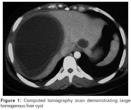
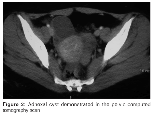
Abdominal ultrasonography showed a cystic lesion measuring 95 × 85 mm in the right lobe of the liver and a cystic lesion of 50 × 40 mm in right adnex. Abdomenopelvic computed tomography (CT) scan with double contrast was performed for further investigation and found a large homogenous cyst in the liver with enhancement of the cyst wall compatible with Type I echinococcal cyst. In addition, a cystic lesion was found in right adnex [Figures 1 and 2].
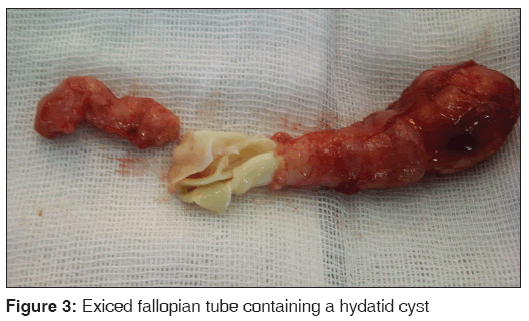
The patient underwent surgery, the liver cyst was excised, the cavity was irrigated with hypertonic saline and omentoplasty was performed. The uterus and adnexa was inspected and a mass was found in right fallopian tube. Salpingectomy was performed and incision of the fallopian tube revealed a cyst containing a clear fluid and white lining typical for germinative layer of echinococcal cyst [Figure 3].
Postoperative course of the patient was uneventful. She was discharged from hospital 2 days later. Oral albendazole 400 mg/d was prescribed. Pathology report confirmed the diagnosis of echinococcal cyst in both liver and fallopian tube.
Discussion
Echinococcosis mostly involves liver and lung. In <20% of patients involvement of other organs happen. Most extrahepatic and extrapulmonary sites are kidney, spleen and peritoneum.[2,3] Hydatid cyst of the fallopian tube is extremely rare, <20 reports could be found in Med Line database. As in other organs of the abdomen and pelvis, hydatid cyst of fallopian tube usually causes nonspecific symptoms due to its compressive effect. Bacterial superinfection and abscess formation may also happen and can be the presenting feature of hydatid cyst. It is highly probable that previously diagnosed pelvic abscess in our patients was a superinfected hydatid cyst in the left adnex.
Preoperative diagnosis is helpful for taking precautions to prevent spillage or considering puncture-aspiration-injection-reaspiration treatment. Serologic test are helpful, but available commercial kits have not high specificity and sensitivity.[4] Imaging plays a major role in diagnosis of hydatid disease; both CT scan and magnetic resonance imaging can assist in diagnosing hydatid disease, especially in the liver.[2,5] Four types of imaging appearances are defined for hydatid cysts: Simple cyst with noninternal architecture, cyst with daughter cysts that shows floating membranes and septa, calcified cyst and cysts with complications like rupture of superinfection.[2,5] Diagnosing hydatid disease in extrahepatic sites by imaging is however difficult since these features have similarities with other benign or malignant lesions.[2] Signs and symptoms of hydatid disease are also nonspecific, so diagnosis of extrahepatic hydatid disease is often suspected by considering epidemiologic factors and prevalence of echinococcosis in patient’s residence area. This diagnosis must be considered especially when a concomitant liver or lung cyst exists. If hydatid cyst involves solely a site that is uncommon for echinococcosis, it will be challenging to make the diagnosis before surgical excision. In our patient, as many reported pelvic hydatid cysts,[6-8] symptoms warranted surgical excision and intraoperative findings led to a diagnosis. Surgical resection is the treatment of choice. Avoiding spillage during surgery is important to prevent recurrence or anaphylaxis. Albendazole is a safe and effective adjuvant therapy and should be administered to minimize recurrence rate after surgery.[9]
Hydatid cyst must be considered in differential diagnosis of every cystic lesion in patients from endemic areas.
References
- McManus DP, Zhang W, Li J, Bartley PB. Echinococcosis. Lancet 2003;362:1295-304.
- Polat P, Kantarci M, Alper F, Suma S, Koruyucu MB, Okur A. Hydatid disease from head to toe. Radiographics 2003;23:475-94.
- Cöl C, Cöl M, Lafçi H. Unusual localizations of hydatid disease. Acta Med Austriaca 2003;30:61-4.
- Zhang W, Wen H, Li J, Lin R, McManus DP. Immunology and immunodiagnosis of cystic echinococcosis: An update. Clin Dev Immunol 2012;2012:101895.
- Marrone G, Crino’ F, Caruso S, Mamone G, Carollo V, Milazzo M, et al. Multidisciplinary imaging of liver hydatidosis. World J Gastroenterol 2012;18:1438-47.
- Uchikova E, Pehlivanov B, Uchikov A, Shipkov C, Poriazova E. A primary ovarian hydatid cyst. Aust N Z J Obstet Gynaecol 2009;49:441-2.
- Alimohamadi S, Dehghan A, Neghab N. Primary bilateral intrapelvic hydatid cyst presenting with adnexal cystic mass: A case report. Acta Med Iran 2011;49:694-6.
- Geramizadeh B. Unusual locations of the hydatid cyst: A review from Iran. Iran J Med Sci 2013;38:2-14.
- Arif SH, Shams-Ul-Bari, Wani NA, Zargar SA, Wani MA, Tabassum R, et al. Albendazole as an adjuvant to the standard surgical management of hydatid cyst liver. Int J Surg 2008;6:448-51.


 The Annals of Medical and Health Sciences Research is a monthly multidisciplinary medical journal.