
Influence of Functional Head Postures on the Dynamic Functional Occlusal Parameters
- *Corresponding Author:
- Dr. Satheesh B Haralur
Department of Prosthodontics, College of Dentistry, King Khalid University, Abha, Kingdom of Saudi-Arabia.
E-mail: hb_satheesh@yahoo.com
Abstract
Background: The dentist utilizes supine position during therapeutic procedures, while the patients assumes extended head posture during mastication. It is critical for the restorative dentist to evaluate and understand the possible effect of change in head posture on occlusal contacts. An understanding of the possible effect will help in better restorative rehabilitation. Aim: The aim of the following study is to evaluate the influence of head postures on the dynamic occlusal parameters. Subjects and Methods: Study group consisted of 50 students from College of Dentistry, King Khalid University. The students were selected randomly from the college student body; they were in the age group of 18‑25 years. The head posture evaluated were supine position, upright sitting position and alert feeding position. The head postures were standardized by goniometer and dynamic occlusal contacts were analyzed with Tscan. The data obtained was subjected to statistical analysis with repeated measures of ANOVA to identify the changes in occlusal contacts. Results: The occlusion time was 1.366 (0.484), 1.226 (0.562) and 0.997 (0.429) s for supine, upright and alert feeding position respectively. Disclusion time was for right lateral movement was 0.872 (0.372), 0.629 (0.290) and 0.831 (0.369) s for corresponding head postures. Left lateral disclusion time was 0.621 (0.274), 0.274 (0.345) s for upright and alert feeding posture. Repeated measures of ANOVA showed statistically significant difference between occlusal contacts in different head postures. Conclusion: Within the limitation of the study, it was concluded that there were significant changes in initial occlusal contact, occlusion and disclusion time at all the head postures evaluated in the study.
Keywords
Occlusal parameters, Head postures, Restorative rehabilitation
Introduction
The efficiency of the masticatory system largely depends on alignment and occlusion of dentition. Pattern of teeth occlusion greatly influence the human vital function such as chewing, swallowing and phonetics. More than static occlusal contacts, dynamic contacts affect the mastication and all other physiological activities. Occlusal contacts are controlled by temporomandibular joint (TMJ), dentition and muscles of mastication. The tooth contact should be along its long axis for the optimum health of the tooth and supporting periodontal structures. Bilateral harmonious contacts will help in the development of orthopedically stable TMJ and maintains the joint health. The mandibular path during the mastication should be devoid of occlusal interferences. Presence of occlusal interference during mastication will result in occlusal instability, increased activity in muscle of mastication.[1] Majority Dental researchers still believe improper occlusal contacts are one of the main etiological factors for the initiation of TMJ disorder.[2]
Head posture is varied according to the physiological and functional activity of the human. These head postures can be divided into an active feeding position posture, upward posture and extended head posture. Incorrect forward head posture is also known to cause the neck, head, shoulder tension and pain along with occlusal changes.[3] Skeletal radiographs, occlusograms were used by the early researchers to establish the positive correlation between head posture and occlusion. Change in head postures will result in a change of mandible position due to stretching and elongation of the muscles attached to it. Routine functional activities such as eating and drinking alter the head posture. The head extends forward by approximately 30° during food consumption; this head posture is known as active feeding posture.[4] This posture shifts the mandible and its closure path anteriorly. The head is extended around 45° during drinking; this will result in the mandible shift posteriorly.[5] All the dental therapeutic procedure’s eventual success depends on its harmony with occlusion. The clinicians most commonly use supine position during restorative procedures, occlusion evaluation and correction. All-important functional occlusal contact during appropriate head position is usually ignored. Existent dental literature on the influence of the functional head posture on the dynamic occlusal contact is minimal. The knowledge of the change in tooth contact during functional head posture will help the dentist during occlusal evaluation and therapeutic procedures. It is critical for dentists to incorporate these changes in occlusion analysis to eliminate the iatrogenic interferences, improve longevity of the restoration and health of all parts of the masticatory system.[6] Therefore, this experimental study was planned with the objective to evaluate the influence of different functional head posture on dynamic occlusal contacts.
Subjects and Methods
Sample
The institutional ethical committee approval was obtained for the study before the initiation. The study methodology was explained, and written consent was obtained from all participants prior to their inclusion in the study. The study sample consisted of 50 students from College of Dentistry, King Khalid University. This study took place in the dental clinics of college of Dentistry, King Khalid University during the first semester of 2013. The King Khalid university college of Dentistry is situated in the southern part of Saudi Arabia. It is one of the largest dental colleges in the kingdom and consists of an undergraduate students ranging from first semester to twelve semester and intern dentists. The student body included the students from all parts of the Kingdom of Saudi Arabia. The student body consisted of 240 students; eighteen students were not interested in participating in the study. The students agreed to participate in the study were screened to the satisfaction of inclusion criteria mentioned in the study. A total of 62 were students identified suitable for study. The simple random sampling procedure was used to select the 50-study sample. All the 62 students university registration numbers were written on the piece of paper in a container, mixed up, randomly selected in the lottery system. This will construct a 95% confidence interval with a Margin of Error of about five. The age group of the subjects included in the study were 18-25 years. The inclusion criteria for the study subjects were: Class I dental occlusion, no posterior missing teeth other than third molars, no previous history of orthodontic treatment or maxillo facial surgery, normal overbite (2-4 mm) and overjet (0-3 mm), no posterior cross bite and no crown/fixed partial dentures. Exclusion criteria was the existence of any signs or symptoms of temporomandibular disorders (TMD).
Head postures
Head postures evaluated in the study were the supine, normal upright sitting and alert feeding position (30° flexion). Rationale of including supine position was due to the fact that most of the dentist uses this chair position during restorative procedures. 30° flexion was included as active feeding head position based on the earlier research findings. Goniometer was used to standardize all the head postures.
Study subjects were made to sit comfortably in the dental chair with lumbar and thoracic spine well-supported by the back of the chair. Shoulder girdle was stabilized by the belt strapped around the dental chair. Supine position was attained by making the subject lay down on his back in the horizontal position with dental chair parallel to the floor. Upright position and 30° head flexure were standardized with the goniometer by following method. For alert feeding position, centre of the goniometer was positioned over the external auditory meatus and proximal arm was held perpendicular to the floor. The distal arm of Goniometer was aligned to base of nares. The subject was asked to flex the head to 30°, the goniometer used to ensure 30° flexion by measuring angle between proximal and distal arms [Figure 1]. Due care was taken not to move the proximal arm during the head movements.

Figure 1: Clinical procedure to measure the 30° flexion
For the upright sitting position, the external auditory meatus was aligned over the shoulder joint. The Patient was requested to occlude over the tongue depressor with the part of it extended outside the mouth. The extended part of the tongue depressor was aligned to be parallel to the floor. Goniometer measurement at 90° was selected [Figure 2]. All the head posture goniometer measurements were done by two researchers, including corresponding author. All the measurements were done under the guidance of the principal author to standardize the goniometer measurements.

Figure 2: Goniometer to standardize the upright head position
Occlusion an evaluation
T scan III (Tekscan Inc., South Boston, USA) was utilized for dynamic occlusal contact evaluation for all three head postures. T scan provides the quantification of occlusal force and a sequence of occlusal contact, a distinct advantage over the conventional occlusal evaluation by articulating paper. All the occlusion evaluation procedures were executed by one researcher under the guidance of the first author.
Occlusion evaluation was initiated after determining the dental arch dimension and intra-oral sensor calibration. Central incisor width was measured with a digital caliper to determine the dental arch dimension. Ideal sensitivity for intra-oral sensor was identified by adjusting the high force-pink areas of contact for a maximum of three graphical displays during trial bite on the sensors. Subjects were guided to centric relation (CR) by the Dawson bimanual method. During occlusal evaluation, the operator guided the mandible to CR and assistant held the articulating ribbon forceps intra-orally.
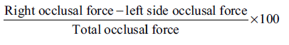
CR mode was selected in the T-scan III toolbar to provide adequate time length and recording sensitivity during CR recording. CR recording provides information on sequential ordering of tooth contacts from an initial tooth contact in CR up to maximum intercuspation. After completion of CR recording other dynamic occlusal contacts such as left lateral, right lateral and protrusive movements were also recorded sequentially. Three successive recordings were performed on all movement to validate the tooth contacts. The occlusion and disclusion time were calculated by observing relative force-time graph. The occlusion time was determined by recording the time lapse from initial tooth contact to maximum intercupation. The disclusion time was calculated at the time required for complete disoccluion from maximum intercuspation in excursive movements. Center of occlusion is shown in T scan graph, centralization of the force is calculated by measuring the distance from this point to the center of the graph. Asymetry of the force was calculated by the following formula:
Statisical analysis
The data obtained was subjected to statistical analysis by SPSS 19 software (Chicago, IL, USA). Repeated measures of ANOVA was utilized for statistical comparison in the variation of occlusal contacts during different head postures.
Ethical consideration
The research ethics committee at college of dentistry, King Khalid University approved the research. The objective and research methodology were explained to all participants and their written consent was obtained before the inclusion in the study.
Results
Occlusion and disocclusion time
Table 1 demonstrates the mean values, one-way ANOVA analysis of occlusion and disclusion times during different head postures.
The occlusion time was 1.366 (0.484) s for the subjects during the supine head posture and 1.226 (0.562) s while the head was in an upright position. The occlusion time was comparatively less (0.997) during the alert feeding head position.
The disclusion time required for right lateral movement was 0.872 (0.372), 0.629 (0.290), 0.831 (0.369) s during supine, upright and 30° forward head postures. The corresponding values for a left lateral diclusion time were 0.862 (0.378) s for supine position. The upright head posture and 30° forward head postures required 0.621 (0.274), 0.274 (0.345) s respectively. The protrusive disclusion time also had similar diclusion time patterns. 1.051 (0.472) s for supine head posture, 0.696 (0.306) s during upright posture and 30° forward head posture had 0.948 (0.451) s.
As shown in the Table 1, there was a significant change in occlusion and disclusion time at different head posture. Hence, these values were subjected to repeated measures of ANOVA to identify the existence of statistically significant variation in occlusal parameters during different head postures. When Mauchley’s test of Sphericity was significant, a Greenhouse Geisser correction of degrees of freedom was used for testing the effect within the subjects. Table 2 presents the Greenhouse-Geisser values for all groups. The analysis showed F values of 18.513, 20.227 and 37.831 for right lateral, left lateral and protrusive disclusion times respectively. P values for all the groups was <0.001. F value for occlusion time was 20.390 with P < 0.05. The result indicated the presence of statistically significant variation between occlusal parameters in different head postures evaluated in the study.
| Occlusion parameters | Head postures | Mean | SD | n | P value | Post-hoc test |
|---|---|---|---|---|---|---|
| Clusion time | Supine (1) | 1.366 | 0.484 | 50 | <0.001 | 1>2>3 |
| Upright (2) | 1.226 | 0.562 | ||||
| 30° forward (3) | 0.997 | 0.429 | ||||
| Right lateral disclusion time | Supine (1) | 0.872 | 0.372 | 50 | <0.001 | 1>3>2 |
| Upright (2) | 0.629 | 0.290 | ||||
| 30° forward (3) | 0.831 | 0.369 | ||||
| Left lateral disclusion time | Supine (1) | 0.862 | 0.378 | 50 | <0.001 | 1>3>2 |
| Upright (2) | 0.621 | 0.274 | ||||
| 30° forward (3) | 0.807 | 0.345 | ||||
| Protrusive disclusoin time | Supine (1) | 1.051 | 0.472 | 50 | <0.001 | 1>3>2 |
| Upright (2) | 0.696 | 0.306 | ||||
| 30° forward (3) | 0.948 | 0.451 | ||||
| SD: Standrad deviation |
SD: Standrad deviation
Table 1: Illustrate the mean values of clusion and disclusion times during different head postures
| Withinsubjects effect | df | Mean square | F value | Significance |
|---|---|---|---|---|
| Clusion time | 1.925 | 1.801 | 2.390 | <0.001 |
| Right lateral | 1.744 | 0.972 | 12.513 | <0.001 |
| disclusion time | ||||
| Left lateral | 1.864 | 0.861 | 20.227 | <0.001 |
| disclusion time | ||||
| Protrusive | 1.816 | 1.842 | 37.831 | <0.001 |
| disclusoin time | ||||
Table 2: Repeated measures of analysis of variance with Greenhouse-Geisser values
Centre of occlusion and asymmetry of occlusion
Centre of Occlusion and Asymmetry of occlusion in supine head position was 4.80 (3.763), 24.821 (19.524) mm. The Upright head posture and 30° forward head posture had a center of occlusion of 4.74 (3.702), 3.96 (3.680) mm respectively. Asymmetry of occlusion for both the head posture was 25.20 (21.27) and 21.16 (18.253) mm respectively. Repeated measures of ANOVA showed an F value of 1.710 for the center of occlusion and 1.192 for asymmetry of occlusion. The respective P values were 0.190 and 0.308. Hence, the statistically insignificant difference was observed among the groups.
Initial occlusal contacts
The initial occlusal contact was different for all groups. 30° forward head posture provided the initial contact majority on anterior teeth 57.8% (30/50) followed by upright 33.3% (16/50) and supine posture 28.9% (14/50). The initial contacts were significantly different among each head posture.
Discussion
Teeth contact patterns play a vital role in determining masticatory efficiency. The success of any restorative treatment is largely dependent on its compliance with good occlusion parameters. Hence, restorations or tooth replacement procedure cannot be initiated nor executed without the proper occlusal evaluation. Accurately identifying and quantifying the occlusal contact, determining their relationships and interference is important in any occlusal evaluation. The dynamic occlusal contacts during the mandibular movement are more important than the static contacts. Knowing the acceptable baseline occlusal parameters are important for the restorative dentist, it will enable him to detect and quantify the unacceptable deviations. The treatment approach like whether to follow confirmative or reorganized approach can be selected depending on the findings. The digital evaluation by T Scan III provides the multiple advantages such as quantification and sequence of occlusal contact. The other important parameters such as occlusion and disclusion time can also be precisely determined.[7]
The studies have shown the influence of head postures on the occlusal contacts.[8] Various head postures are utilized for better visibility and accessibility during routine dental treatments. The supine and upright head postures are routinely used during diagnostic occlusion evaluation to final restorations. The head is flexed by average 30° during the eating, existence of substantial change in occlusal contact between these head posture may be detrimental to the teeth, restorations and their supporting structures. Hence, the dynamic tooth contact changes during these head postures are critical for a dentist to understand.
Identifying the initial tooth contact (CR), premature contacts and slide from CR to maximum intercuspation is important for the health of the teeth, supporting structure and TMJ.[9] Researchers observed the properly positioned condyles in the articular fossae during CR has the least muscle activity.[10] The results of the study showed the change in initial tooth contact position in all three head positions. An alert feeding position with 30° forward head posture had the predominantly anterior initial contacts, while supine position only had few initial contacts on anterior segment. The change on initial contact can be justified by the observation of Preiskel [11] and Goldstein et al.[12] They demonstrated the relation between head position and mandibular position. The literature suggests [13] the contraction of mandibular elevator muscles during alert feeding position shift the path of mandibular closure slightly anterior to upright head position. Hence, the anterior teeth receive the heavy contact in this head posture than upright position.
The time required by the anterior guidance to disengage all posterior teeth in protrusive and lateral movements is known as disclusion time. The disclusion time more than 1.39 s reported to initiate the elevated muscular contraction in masseter and temporalis.[14,15] The EMG study showed the resting state muscular activity was observed in disclusion time <0.5 s. The prolonged disclusion time is also reported to be the etiological factor for myofunctional pain dysfunction syndrome.[15] Disclusion time during supine position was 0.872 (0.372), 0.862 (0.378), 1.051 (0.472) s for left lateral, right lateral and protrusive disclusion. The alert feeding position had a disclusion time of 0.831 (0.369), 0.274 (0.345) and 0.948 (0.451) s for similar mandibular movements. 0.629 (0.290), 0.621 (0.274), 0.696 (0.306) s were required for the disclusion during upright head posture. The statistically significant (P < 0.5) differences were observed at disclusion time among all three head postures.
The occlusion time is the time elapsed between the initial contacts to the maximum static intercuspation of teeth. This time ideally is as short as possible, longer occlusion time is detrimental in nature to the masticatory system.[16] According to the results of the study, there were significant changes were found in occlusion time and disclusion times among different head postures. Occlusion time was longest in supine head position 10.366 (0.484) s, while alert feeding position had the least occlusion time of 0.997 (0.429) s. The mandible is located 2-4 mm below the maximum intercuspation in upright head posture, while during head flexion or extension result in associated elevator muscle contraction. The change in mandibular postural position might be the cause of changes in occlusion and disclusion time.
The center of occlusion indicates the balance in occlusion. Studies indicate the balance of the force in the center of the arch bilaterally and Antero-posteriorly in the region on the first molar. Studies have [17] demonstrated the asymmetry of force is higher in the patient with TMD disorders than the normal subjects. Hence, both centers of occlusion and asymmetry of occlusion are considered as important diagnostic criteria. The results of the study showed the Asymmetry of force was observed more in the supine position compared other head postures.
Within the limitation of the Study, it can be concluded that there is a strong correlation between head postures and dynamic occlual contacts. The dentists should consider evaluating occlusion in functional head postures, rather than only in a supine head position or upright head posture. The dynamic tooth contact during functional head posture is to be evaluated, corrected and monitored for the long-term success of restorations.
Conclusion
The dynamic occlusal contacts during the functional head postures should be accounted during the diagnostic and therapeutic procedures. This study demonstrated the change in the initial contact between supine, upright and active feeding head postures. The occlusion and disclusion times were also significantly different in all head postures evaluated. The asymmetry of force was found to be highest in the supine position. During the management of occlusion during the restorative procedures and TMD these changes should ideally be taken into consideration.
Acknowledgment
The study was self-financed; the authors would like to thank King Khalid University for providing the facility to evaluate the participants.
Source of Support: Nil.
Conflict of Interest: None declared.
References
- Kerstein RB, Radke J. Masseter and temporalis excursive hyperactivity decreased by measured anterior guidance development. Cranio 2012;30:243-54.
- Reinhardt R, Tremel T, Wehrbein H, Reinhardt W. The unilateral chewing phenomenon, occlusion, and TMD. Cranio 2006;24:166-70.
- Weon JH, Oh JS, Cynn HS, Kim YW, Kwon OY, Yi CH. Influence of forward head posture on scapular upward rotators during isometric shoulder flexion. J Bodyw Mov Ther 2010;14:367-74.
- Mohl N. Head posture and its role in occlusion. Int J Orthod 1977;15:6-14.
- Ohmure H, Miyawaki S, Nagata J, Ikeda K, Yamasaki K, Al-Kalaly A. Influence of forward head posture on condylar position. J Oral Rehabil 2008;35:795-800.
- Koidis PT, Novak M, Magiliotou MC, Burch JG. Influence of postural position on occlusal contact strain patterns. J Dent Res 1986;65:189.
- Ma FF, Hu XL, Li JH, Lin Y. Normal occlusion study: Using T-Scan III occlusal analysis system. Zhonghua Kou Qiang Yi Xue Za Zhi 2013;48:363-7.
- Makofsky HW. The influence of forward head posture on dental occlusion. Cranio 2000;18:30-9.
- Troeltzsch M, Troeltzsch M, Cronin RJ, Brodine AH, Frankenberger R, Messlinger K. Prevalence and association of headaches, temporomandibular joint disorders, and occlusal interferences. J Prosthet Dent 2011;105:410-7.
- Ramfjord SP. Bruxism, a clinical and electromyographic study. J Am Dent Assoc 1961;62:21-44.
- Preiskel HW. Some observations on the postural position of the mandible. J Prosthet Dent 1965;15:625-33.
- Goldstein DF, Kraus SL, Williams WB, Glasheen-Wray M. Influence of cervical posture on mandibular movement. J Prosthet Dent 1984;52:421-6.
- McLean LF, Brenman HS, Friedman MG. Effects of changing body position on dental occlusion. J Dent Res 1973;52:1041-5.
- Kerstein RB, Radke J. The effect of disclusion time reduction on maximal clench muscle activity levels. Cranio 2006;24:156-65.
- Michelotti A, Cioffi I, Festa P, Scala G, Farella M. Oral parafunctions as risk factors for diagnostic TMD subgroups. J Oral Rehabil 2010;37:157-62.
- Weffort SY, de Fantini SM. Condylar displacement between centric relation and maximum intercuspation in symptomatic and asymptomatic individuals. Angle Orthod 2010;80:835-42.
- Yamada R, Ogawa T, Koyano K. The effect of head posture on direction and stability of mandibular closing movement. J Oral Rehabil 1999;26:511-20.


 The Annals of Medical and Health Sciences Research is a monthly multidisciplinary medical journal.