Molecular Evaluation and Seroprevalence of Toxoplasmosis in Pregnant Women in Fars province, Southern Iran
2 Department of Basic Sciences in Infectious Diseases Research Center, Shiraz University of Medical Sciences, Shiraz, Iran, Email: sarkarib@sums.ac.ir
3 Department of Parasitology and Mycology, Faculty of Medicine, Zahedan University of Medical Sciences, Zahedan, Iran, Email: abdolahi.khabisi_s@gmail.com
Citation: Norouzi Larki Y, Sarkari B, Asgari Q, Abdolahi Khabisi S. Molecular Evaluation and Seroprevalence of Toxoplasmosis in Pregnant Women in Fars province, Southern Iran. Ann Med Health Sci Res. 2017; 7:16-19.
This open-access article is distributed under the terms of the Creative Commons Attribution Non-Commercial License (CC BY-NC) (http://creativecommons.org/licenses/by-nc/4.0/), which permits reuse, distribution and reproduction of the article, provided that the original work is properly cited and the reuse is restricted to noncommercial purposes. For commercial reuse, contact reprints@pulsus.com
Abstract
Background: Importance of Toxoplasma gondii for humans refers mainly to primary infection in pregnant women and also infection in immunocompromised individuals. Aim: The current study aimed to determine the seroprevalence of toxoplasmosis in pregnant women in Fars province, southern Iran, and to find out the chronic and acute cases of toxoplasmosis in this population with molecular and serological methods. Subjects and Methods: Blood samples were taken from 2000 pregnant women, admitted to Shiraz university-affiliated hospitals in 2014 and serum and buffy coat were separated. Data such as age, number of pregnancy, pregnancy age and place of resident were recorded for each participant. Sera samples were evaluated for anti-Toxoplasma IgG and IgM, using a commercial ELISA kit. Moreover, the seropositive cases were evaluated by PCR to amplify a 529bp gene of Toxoplasma gondii. Results: Anti-Toxoplasma antibodies were detected in sera of 177 (8.9%) of cases. From these, 172 (8.6%) were seropositive only for IgG, 4 (0.2%) were seropositive only for IgM and 1 (0.05%) were positive for both IgG and IgM. PCR detected Toxoplasma DNA in buffy coat of 15 out of 177 (8.5%) of the seropositive subjects, two of them were IgM positive and the remaining 13 were among IgG seropositive cases. Conclusion: Findings of this study demonstrated a relatively low rate of seropositivity for toxoplasmosis in pregnant women in the area. This means that a high percentage of pregnant women are at risk of acquiring toxoplasmosis during their pregnancy and subsequently, transmission of the infection to their fetus
Keywords
Seroprevalence, Molecular evaluation, Toxoplasmosis, Pregnant women, Southern Iran
Introduction
Toxoplasma gondii is a cosmopolitan zoonotic protozoan which can infect many of warm blooded animals such as birds, mammals, and human.[1] Rate of human infection in different areas of the world is different due to variation in nutritional diets of the people, weather condition and reservoir hosts.
Infections with toxoplasmosis usually cause a mild disease in immunocompetent individuals and severe and life threatening infection in immunocompromised patient. The most serious manifestation of the disease can be seen in congenital toxoplasmosis, resulting from vertical transmission of the parasite during pregnancy.[2-4] Seroprevalence of toxoplasmosis in women at childbearing age varies in different geographical areas of Iran and ranged from 5 up to 70%. [5]
prevalence rate of 55.5% has been reported form toxoplasmosis among pregnant women in north of Iran while this rate has been reported to be 29.3% in Khuzestan, south of Iran, with a different weather condition.[6-7] Fallah et al. reported a seroprevalence of 33.5% for Toxoplasma in pregnant women in 2004 in Hamedan, western Iran.[8] A seroprevalence rate of 34% was reported for anti-Toxoplasma antibodies in pregnant women from Qazvin, in northwest of Iran.[9] Mostafavi et al. documented a seroprevalence of 47% in pregnant women in Isfahan, central Iran, and Davami et al. reported a seroprevalence of 15% for toxoplasmosis for women who referred to clinical laboratories for pre-marriage testing.[10,11]
It has been documented that Toxoplasma DNA can be detected in the buffy coat of individual during acute toxoplasmosis.[12] This might be a very useful approach for diagnosis of acute toxoplasmosis in pregnant women, where defining the acute infection in crucial for implementation of appropriate therapy and prevention measurements.
The current study aimed to determine the seroprevalence of toxoplasmosis in pregnant women in Fars province, southern Iran, and to find out the chronic and acute cases of toxoplasmosis in this population with molecular and serological methods.
Subjects and Methods
Study population
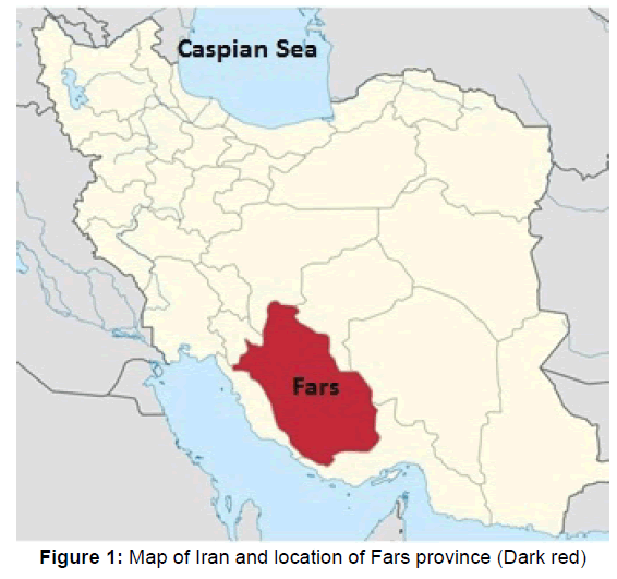
The current study was conducted in 2014 in Fars province, southern Iran [Figure 1]. After getting approval from the ethics committee of Shiraz University of Medical Sciences, blood samples were taken from 2000 pregnant women admitted to Shiraz university-affiliated hospitals. Sample size was calculated based on the prevalence of toxoplasmosis in the region, using SPSS software (version 18, SPSS, Chicago). Demographic features and also number of pregnancy, pregnancy age and place of resident were recorded for each participant.
Serological tests
Sera were obtained from the whole blood of each participant. Moreover, buffy coat was obtained from each blood sample for subsequent molecular study. Samples were transferred to the serology laboratory at department of parasitology and mycology in Shiraz University of Medical Sciences, Shiraz, Iran, and kept at -20°C until use. All of sera samples were evaluated for anti-Toxoplasma IgG and IgM antibodies, using a commercial enzyme immunoassay kit (Pishtaz Teb Diagnostics, Tehran, Iran).
Molecular analysis
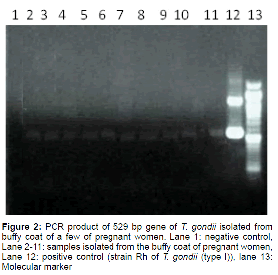
The seropositive cases (either IgG or IgM) were evaluated by PCR to detect Toxoplasma DNA in the buffy coats. DNA was extracted from the buffy coat of each sample, using phenol chloroform method. PCR was performed to amplify a 529 bp gene of T. gondii, as described by Edvinsson et al. using each of two primers, TOXOF CAGGGAGGAAGACGAAAGTTG and TOXOR CAGACACAGTGCATCTGGATT.[13]
Statistical analysis of the data
Collected data were analyzed by SPSS software (version 18, SPSS, Chicago). Fisher exact test and chi-square were used to compare the seroprevalence values related to the features of the subjects.
Results
The pregnant women being studied were aged from 15 to 45 years old with mean age of 27.4 years. Most of the participants (35.4%) were aged 25-30 years old and the lowest (1.2%) were in the age group of 40 or higher. Most of pregnant women (57.3%) were in the second trimester of their pregnancy. Anti T. gondii antibodies were detected in the sera of 177 out of 2000 pregnant women, corresponding to an overall seroprevalence of 8.9% in this population. Of these, 172 (8.6%) were seropositive for only IgG, 4 (0.2%) were seropositive for only IgM and 1 (0.05%) was positive for both IgG and IgM.
Most of the seropositive cases (36.2%) were younger than 25 years old and the lowest (1.1%) were aged higher than 40 years old. Chi square test was used to find out the statistical association between seropositivity with number of pregnancy, trimesters of pregnancy, and age of the participants. There were a significant association between seropositivity and number and also trimesters of pregnancy of the participants (p<0.01). Most of IgM positive cases (0.3%) were seen in the age group of 15-30 years old whereas pregnant women aged 30-35 years had the lowest rate of IgM seropositivity. Correlation between IgM positivity and number of pregnancy or trimester were not statistically significant (p=0.18). All of seropositive cases were tested for the presence of T. gondii DNA in their buffy coat samples. Molecular study revealed a 529bp band of T. gondii in peripheral blood of 15 out of 177 (8.5%) seropositive pregnant women [Figure 2]. Two of these cases were IgM positive and the remaining 13 cases were among the IgG seropositive cases. Table 1 shows the demographic features of pregnant women and relative seropositivity to T. gondii in this study.
| Characteristics | Frequency | Percent (%) | Positive for anti-Toxoplasma antibodies (either IgG or IgM) | P Value | |
|---|---|---|---|---|---|
| No | % | ||||
| Age Group (Year) | |||||
| <25 | 689 | 34.5 | 64 | 9.3 | 0.16 |
| 25-30 | 707 | 35.4 | 54 | 7.6 | |
| 31-35 | 448 | 22.4 | 38 | 8.5 | |
| 36-40 | 132 | 6.6 | 19 | 14.4 | |
| >40 | 24 | 1.2 | 2 | 8.3 | |
| Pregnancy Age | |||||
| First Trimester | 23 | 1.2 | 6 | 26.1 | < 0.01 |
| Second Trimester | 1146 | 57.3 | 139 | 12.1 | |
| Third Trimester | 831 | 41.6 | 32 | 3.9 | |
| Pregnancy Term | |||||
| First | 1030 | 51.5 | 109 | 10.6 | < 0.01 |
| Second | 885 | 44.3 | 56 | 6.3 | |
| Third | 77 | 3.9 | 11 | 14.3 | |
| Fourth | 7 | 0.4 | 1 | 14.3 | |
| Fifth | 1 | 0.1 | 0 | 0 | |
Table 1: Demographic features of pregnant women and relative seropositivity to T. gondii in Fars province, southern Iran
Discussion
Toxoplasmosis is a common parasitic infection of human and animals throughout the world, including Iran.[1,4,10,14] The overall seroprevalence rate of toxoplasmosis among the general population in Iran was reported to be 39.3%.[5]
In the current study seropositive rate of toxoplasmosis in pregnant women was found to be 8.8%, in southern Iran. This rate of seroprevalence in pregnant women is more or less similar to the rates reported in other studies conducted on child bearing and pregnant women in southern part of the country but lower than those reported from north part of the country.[6,7,9,15] Rate of Toxoplasma infection in north part of the country, where the temperature is ambient for survival of Toxoplasma oocyst excreted by the cat, is higher in comparison to the southern and western part of the country.[6,7,16]
Keeping cats and dogs as pets are not so common in Fars province in Iran. However, the number of stray cats and dogs are significant and stray cats, in this case, pose a major threat to human health through their role as reservoirs of Toxoplasma.
Consumption of contaminated vegetables might be the main source of Toxoplasma infection in pregnant women since in Iran women are usually clean and wash the vegetables at home and they may contaminate themselves while cleaning the vegetables. However, high seroprevalence of toxoplasmosis in meat producing animals in the region indicates that transmission may be occurring through consumption of undercooked meats of farm animals, as likely as contaminated vegetables.[17,18]
In the current study, rate of seropositivity was increased with pregnancy number where seropositivity was higher in multigravidae than in primigravidae. This is probably related to the age of the subjects rather than number of pregnancy, since the rate of seropositivity to Toxoplasma usually increase with the age.
The presence of IgM anti-Toxoplasma antibodies reflects the risk of transplacental transmission. This is not always the case since IgM may persist as long as one year in sera of toxoplasmic patients. Therefore, other serological methods might be used to find out the estimate time of infection. Detection of Toxoplasma DNA in the buffy coat of the seropositive individuals may be related to acute infection. In a recent study in Fars province, south of Iran, 1500 cases of blood donors were evaluated for anti- Toxoplasma antibodies and 5.4% of the donors were seropositive for IgM and 1.6% for both IgG and IgM. Toxoplasma DNA has been detected in blood samples of two of IgM positive cases of blood donors but not in the buffy coat of the rest of seropositive cases.[14] In another study conducted on patients undergoing chemotherapy for malignancies in the Bushehr Province, Southwest Iran, T. gondii DNA was detected in the buffy coats of two out of 86 (2.3%) cases.[19] In the current study IgM anti- Toxoplasma antibody were detected in the sera of 5 (0.25%) of pregnant women while PCR detected Toxoplasma DNA in the buffy coat of 15 (0.75%) of the seropositive subjects, two of them were IgM positive and the remaining 13 were among IgG seropositive cases. Based on these findings, detection of Toxoplasma DNA in the buffy coat may not necessary indicate an acute infection. Moreover, presence of Toxoplasma DNA in the blood of IgM negative cases might be related to the false negativity of IgM result.
Taken together findings of this study showed that a low number of pregnant women in Fars province, southern Iran, are seropositive for toxoplasmosis. This indicates that high percentages of pregnant women are at risk of acquiring toxoplasmosis during their pregnancy and subsequent transmission of the infection to their fetus. Furthermore, in this study, molecular findings had no relationship with acute toxoplasmosis in studied cases. Further study, with large number of IgM positive cases, is needed to explore the value of PCR detection of Toxoplasma DNA in buffy coat for diagnosis of acute toxoplasmosis.
Conflict of Interest
The authors declare that there is no conflict of interests regarding the publication of this paper.
REFERENCES
- Robert-Gangneux F, Darde ML. Epidemiology of and diagnostic strategies for toxoplasmosis. Clin Microbiol Rev. 2012;25:264-296.
- Torgerson PR, Mastroiacovo P. The global burden of congenital toxoplasmosis: A systematic review. Bull World Health Organ. 2013; 91:501-508.
- Sarkari B, Abdolahi Khabisi S. Severe congenital toxoplasmosis: A case report and strain characterization. Case Rep Infect Dis. 2015; 2015:851085.
- Asgari Q, Fekri M, Monabati A, Kalantary M, Mohammadpour I, Motazedian MH, et al. Molecular genotyping of Toxoplasma gondii in human spontaneous aborted fetuses in Shiraz, southern Iran. Iran J Public Health. 2013;42:620-625.
- Daryani A, Sarvi S, Aarabi M, Mizani A, Ahmadpour E, Shokri A, et al. Seroprevalence of Toxoplasma gondii in the Iranian general population: A systematic review and meta-analysis. Acta Trop. 2014;137:185-194.
- Sharif M, Daryani A, Ebrahimnejad Z, Gholami S, Ahmadpour E, Borhani S, et al. Seroprevalence of anti-Toxoplasma IgG and IgM among individuals who were referred to medical laboratories in Mazandaran province, northern Iran. J Infect Public Health. 2016; 9:75-80.
- Yad MJ, Jomehzadeh N, Sameri MJ, Noorshahi N. Seroprevalence of Anti-Toxoplasma gondii antibodies among pregnant woman in South Khuzestan, Iran. Jundishapur J Microbiol. 2014;7:e9998.
- Fallah M, Rabiee S, Matini M, Taherkhani H. Seroepidemiology of toxoplasmosis in primigravida women in Hamadan, Islamic Republic of Iran, 2004. East Mediterr Health J. 2008;14:163-171.
- Hashemi HJ, Saraei M. Seroprevalence of Toxoplasma gondii in unmarried women in Qazvin, Islamic Republic of Iran. East Mediterr Health J. 2010;16:24-28.
- Davami MH, Pourahamd M, Jahromi AR, Tadayon SM. Toxoplasma seroepidemiology in women who intend to marry in Jahrom, Islamic Republic of Iran. East Mediterr Health J. 2014;19:S71-S75.
- Mostafavi N, Ataei B, Nokhodian Z, Monfared LJ, Yaran M, et al. Toxoplasma gondii infection in women of childbearing age of Isfahan, Iran: A population-based study. Adv Biomed Res. 2012;1:60.
- Kompalic-Cristo A, Frotta C, Suarez-Mutis M, Fernandes O, Britto C. Evaluation of a real-time PCR assay based on the repetitive B1 gene for the detection of Toxoplasma gondii in human peripheral blood. Parasitol Res. 2007;101:619-625.
- Edvinsson B, Jalal S, Nord CE, Pedersen BS, Evengard B. Toxoplasmosis ESGo: DNA extraction and PCR assays for detection of Toxoplasma gondii. APMIS. 2004; 112:342-348.
- Sarkari B, Shafiei R, Zare M, Sohrabpour S, Kasraian L. Seroprevalence and molecular diagnosis of Toxoplasma gondii infection among blood donors in southern Iran. J Infect Dev Ctries. 2014;8:543-547.
- Khanaliha K, Motazedian M, Sarkari B, Bandehpour M, Sharifnia Z, et al. Expression and purification of P43 Toxoplasma gondii surface antigen. Iran J Parasitol. 2012;7:48-53.
- Sharbatkhori M, Moghaddam DY, Pagheh AS, Mohammadi R, Mofidi HH, et al. Seroprevalence of Toxoplasma gondii infections in pregnant women in Gorgan city, Golestan Province, Northern Iran-2012. Iran J Parasitol. 2014;9:181-187.
- Asgari Q, Sarnevesht J, Kalantari M, Sadat SJ, Motazedian MH, et al. Molecular survey of Toxoplasma infection in sheep and goat from Fars province, Southern Iran. Trop Anim Health Prod. 2011;43:389-392.
- Sarkari B, Asgari Q, Bagherian N, Esfahani AS, Kalantari M, et al. Molecular and serological evaluation of Toxoplasma gondii infection in reared turkeys in Fars Province, Iran. Jundishapur J Microbiol. 2014;7:e11598.
- Barazesh A, Sarkari B, Sisakht MF, Khabisi AS, Nikbakht R, et al. Seroprevalence and molecular evaluation of toxoplasmosis in patients undergoing chemotherapy for malignancies in the Bushehr Province, Southwest Iran. Jundishapur J Microbiol. 2016;17:e35410.


 The Annals of Medical and Health Sciences Research is a monthly multidisciplinary medical journal.