Prevalence of Meniscal Extrusion and associated Knee Findings Detected by MRI in Patients with a History of Knee Trauma
Media S Ibrahim1* and Sameeah A Rashid2
1Department of Radiology, Rizgary Teaching Hospital, Erbil, Iraq
2Department of Surgery, College of Medicine, Hawler Medical University, Erbil, Iraq
- *Corresponding Author:
- Media S Ibrahim
Department of Radiology
Rizgary Teaching Hospital
Erbil, Iraq
Tel: 07702213003
E-mail: media.salih83@gmail.com
Citation: Ibrahim MS, et al. Prevalence of Meniscal Extrusion and associated Knee Findings Detected by MRI in Patients with a History of Knee Trauma. Ann Med Health Sci Res. 2020;10: 870-876.
This is an open access article distributed under the terms of the Creative Commons Attribution‑NonCommercial‑ShareAlike 3.0 License, which allows others to remix, tweak, and build upon the work non‑commercially, as long as the author is credited and the new creations are licensed under the identical terms.
Abstract
Background and objectives: Meniscal extrusion is defined as the condition in which the meniscus is located outside the margin of the knee joint. This condition can lead to knee osteoarthritis, meniscal degeneration, and meniscal tears. Meniscal extrusion is broadly diagnosed through magnetic resonance imaging. The present study aimed to examine the prevalence of meniscal extrusion and the associated knee joint abnormalities in cases of trauma. Methods: The present cross-sectional study consisted of 122 cases (62 cases with knee injuries caused by falling on ground and 60 as a result of football playing) whom been referred to MR unit to undergo knee exam referred in the period from January to July 2019. A search for meniscus extrusion was performed, and once identified then checking for associated findings was performed. Results: The study group mean age was 31.77 years. Most of the cases (77%) were males. About one third of the cases (30.3) had meniscal extrusion, and almost half of them (48.4%) had meniscal tear. Meniscal root tear and complex tear of posterior horn of medial meniscus was the most frequent meniscal tear. About half of the cases (49.2%) had joint effusion. In both study subgroup, meniscal extrusion was significantly found in the medial meniscus, and it was significantly associated with medial meniscus tear, types of meniscal tear, and joint effusion (p<0.05). Age had a significant association with meniscal extrusion in the subgroup of trauma caused by falling on ground (p<0.05), the two groups were significantly different in terms of the meniscal extrusion distance (p<0.05). Conclusion: Medial meniscus and medial meniscus tear are the most significant risk factors for meniscal extrusion. Meniscal extrusion distance was shorter in individuals with knee trauma caused by football playing compared to those with knee injuries resulted from falling on ground.
Keywords
Meniscal extrusion; Joint abnormalities; Magnetic Resonance Imaging (MRI); Knee trauma
Introduction
The menisci refer to two fibro cartilaginous discs that are located laterally and medially between the surfaces of tibia and femur in the cavity of the knee joint. [1] The menisci are highly significant because they stabilize the knee joint, reduce friction and shock during movement, and distribute the body weight across the knee joint. [2,3] According to the results of biomechanical studies, the menisci transmit a minimum of 50% of the knee joint compressive load. [4] As a result, any change in the integrity and normal place of the menisci can lead to negative impacts on these important functions. As revealed by the results obtained from magnetic resonance imaging (MRI) tears or destruction of the menisci are quite common in the general population and rise with age. [5,6]
One of the destructions in the menisci is labeled as meniscal extrusion (also referred to as meniscal subluxation) which is defined as a condition in which the peripheral border of the meniscus is remarkably located outside the margin of the knee joint. [7,8] Meniscal extrusion has been reported to be associated with the presence of knee osteoarthritis (OA), meniscal degeneration, and meniscal tears. [9-12]
A study has shown that women experience meniscal extrusion more than men, which is attributed to the higher laxity of special knee structures like collateral ligaments in women compared to men. [13] In addition to female sex, body mass index in the ranges of overweight and obesity can also cause meniscal extrusion to develop. [11] Furthermore, studies have indicated association between extrusion of the meniscus, osteoarthritis, and joint effusion. [14,15] Also, meniscal extrusion has been reported after knee trauma. [16,17]
According to what was mentioned above and given the significance of diagnosing meniscal extrusion and the associated knee abnormalities, the present study was aimed at determining the prevalence of meniscal extrusion and the associated knee joint abnormalities in two different subgroups of patients (falling on ground and football player), and making a comparison between these two subgroup findings.
Methods and Materials
Study design, setting, and sample
The present cross-sectional study was carried out on 122 cases with a history of knee trauma over a period of 6 months from January to July 2019. The patients were selected from those who referred to Rizgary Teaching Hospital in Erbil, the Kurdistan Region of Iraq. The study sample was chosen by a convenience sampling method. Patients with degenerative changes, claustrophobic patients, patients with contraindication to do MRI and failure to obtain appropriate image and sequences were crossed out of the study.
Procedures
The MR studies were performed on a 1.5 -T unit (Aera, Siemens, Erlangen, Germany), and a knee coil was used in all cases. The examination protocol included sagittal T1 weighted turbo spinecho sequence (TR range/TE range, 400-500/15-20), sagittal T2-weighted turbo spin-echo sequence (TR range/TE range, 2500-3000/74-130), axial, sagittal and coronal proton density turbo-spin echo sequences with fat suppression (TR range/TE range, 2000-2500/40-80) with a 3-mm section thickness and a 0.3 mm gap. The field of view was 20 cm, with a matrix size of 256 × 256.
Image analysis
Two radiologists evaluated the MR images of a total of 122 cases. The study group was classified as sport injury (60 athletes, football player; all men) and trauma due to fall on ground (62 patients, 34 males, 28 females) that has been referred to MR unit from the orthopedic department.
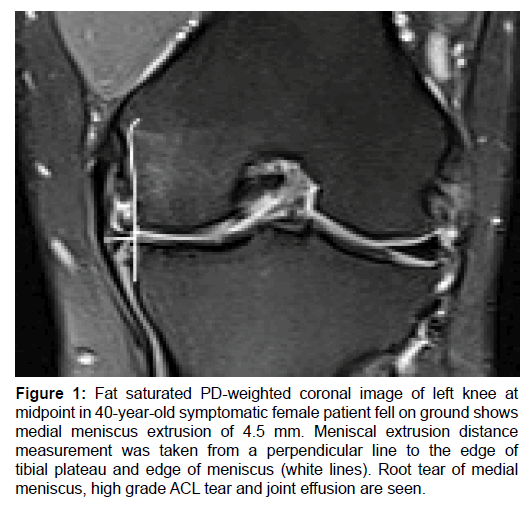
To look for meniscus extrusion and its measurement, coronal plane was used as a reference, based on the technique described by Breitenseher et al. [17] Meniscus extrusion was defined as a distance of 3 mm or more between the peripheral edge of the meniscus and the central portion of the tibial plateau as measured in coronal plane [Figure 1]. A distance of less than 3 mm was not considered as meniscal extrusion. A search for other types of knee joint injuries was also made.
Figure 1: Fat saturated PD-weighted coronal image of left knee at midpoint in 40-year-old symptomatic female patient fell on ground shows medial meniscus extrusion of 4.5 mm. Meniscal extrusion distance measurement was taken from a perpendicular line to the edge of tibial plateau and edge of meniscus (white lines). Root tear of medial meniscus, high grade ACL tear and joint effusion are seen.
Meniscus tear was considered when an internal meniscal signal intensity is identified extending to the articular surfac. Meniscal extrusion was correlated with meniscal tear, joint effusion, and other knee joint injuries (like collateral ligaments, and PCL tear). Cases of mild, moderate and large joint effusion were enrolled in our study, while cases of small physiologic joint fluid were not considered in our study. Both low-grade and high-grade ACL tears were considered as tear in our study.
Statistical analysis
The collected data from the conducted examinations for each patient were recorded using a special questionnaire designed for diagnosis, and they were later analyzed using SPSS version 23 and a Fisher’s exact test was used to assess the significance of the associations between meniscal extrusion and knee joint abnormalities.
Ethical considerations
The study protocol was approved by the Ethical and Scientific Committee of Kurdistan Board for Medical Specialties. The purpose of the study was explained to each patient, and a verbal informed consent was obtained from them prior to their inclusion in the study.
Results
The final number of our study group was 122 cases, with mean age of 31.77 (±9.18) years, most (73.8%) were adults (25-64 years), 23.8% were young (15-24 years), and 2.5% were children (<14 years). Of the 122 patients, 94 (77%) were males, and 28 (23%) females. The prevalence of meniscus extrusion was 30.3% (37 cases), and it was significantly higher in the medial than the lateral meniscus (P value of 0.001). A significant association was found between meniscus extrusion and meniscus tear, type of the tear, joint effusion and PCL tear, but no significant association was found to ACL, MCL and LCL tear. (The frequency of the mentioned data and their statistical correlation are shown in Tables 1 and 2.
| Frequency | Percentage | Frequency | Percentage | ||
|---|---|---|---|---|---|
| Meniscal Extrusion | Meniscal tear | ||||
| Yes | 37 | 30.3 | Yes | 59 | 48.4 |
| No | 85 | 69.7 | No | 63 | 51.6 |
| Total | 122 | 100.0 | Total | 122 | 100.0 |
| Lateral meniscus extrusion | Medial Meniscal tear | ||||
| Yes | 1 | .8 | Yes | 56 | 45.9 |
| No | 121 | 99.2 | No | 66 | 54.1 |
| Total | 122 | 100.0 | Total | 122 | 100.0 |
| Medial meniscus extrusion | Lateral meniscal tear | ||||
| Yes | 36 | 29.5 | Yes | 3 | 2.5 |
| No | 86 | 70.5 | No | 119 | 97.5 |
| Total | 122 | 100.0 | Total | 122 | 100.0 |
| Joint effusion | ACL tear | ||||
| Yes | 60 | 49.2 | No | 59 | 48.4 |
| No | 62 | 50.8 | High grade | 31 | 25.4 |
| Total | 122 | 100.0 | Low grade | 32 | 26.2 |
| Total | 122 | 100.0 | |||
| PCL tear | MCL tear | ||||
| No | 110 | 90.2 | Yes | 5 | 4.1 |
| High grade | 1 | .8 | No | 117 | 95.9 |
| Low grade | 11 | 9.0 | Total | 122 | 100.0 |
| Total | 122 | 100.0 | |||
| LCL tear | |||||
| Yes | 1 | .8 | |||
| No | 121 | 99.2 | |||
| Total | 122 | 100.0 | |||
MCL: Medial Collateral Ligament, LCL: Lateral Collateral Ligament.
Table 1: The frequency and percentage of meniscal extrusion and associated knee findings.
| Extrusion | |||||
|---|---|---|---|---|---|
| Yes | No | Total | P-value | ||
| Medial meniscus extrusion | Yes | 36 (100.0) | 0.001 | ||
| No | 1 (1.2) | 85 (98.8) | 86 (100.0) | ||
| Total | 37 (30.3) | 85 (69.7) | 122 (100.0) | ||
| Lateral meniscus Extrusion | Yes | 1 (100.0) | 0 (0.0) | 1 (100.0) | 0.30 |
| No | 36 (29.8) | 85 (70.2) | 121 (100.0) | ||
| Total | 37 (30.3) | 85 (69.7) | 122 (100.0) | ||
| Meniscal tear | Yes | 35 (59.3) | 24 (40.7) | 59 (100.0) | 0.001 |
| No | 2 (3.2) | 61 (96.8) | 63 (100.0) | ||
| Total | 37 (30.3) | 85 (69.7) | 122 (100.0) | ||
| Medial meniscus tear | Yes | 34 (60.7) | 22 (39.3) | 56 (100.0) | 0.001 |
| No | 3 (4.5) | 63 (95.5) | 66 (100.0) | ||
| Total | 37 (30.3) | 85 (69.7) | 122 (100.0) | ||
| Lateral meniscus tear | Yes | 1 (33.3) | 2 (66.7) | 3 (100.0) | 1.00 |
| No | 36 (30.3) | 83 (69.7) | 119 (100.0) | ||
| Total | 37 (30.3) | 85 (69.7) | 122 (100.0) | ||
| Joint effusion | Yes | 27 (45.0) | 33 (55.0) | 60 (100.0) | 0.001 |
| No | 10 (16.1) | 52 (83.9) | 62 (100.0) | ||
| Total | 37 (30.3) | 85 (69.7) | 122 (100.0) | ||
| ACL tear | No | 21 (35.6) | 38 (64.4) | 59 (100.0) | 0.279 |
| High grade | 6 (19.4) | 25 (80.6) | 31 (100.0) | ||
| Low grade | 10 (31.3) | 22 (68.8) | 32 (100.0) | ||
| Total | 37 (30.3) | 85 (69.7) | 122 (100.0) | ||
| PCL tear | No | 30 (27.3) | 80 (72.7) | 110 (100.0) | 0.033 |
| High grade | 0 (0.0) | 1 (100.0) | 1 (100.0) | ||
| Low grade | 7 (63.6) | 4 (36.4) | 11 (100.0) | ||
| Total | 37 (30.3) | 85 (69.7) | 122 (100.0) | ||
| MCL tear | Yes | 3 (60.0) | 2 (40.0) | 5 (100.0) | 0.163 |
| No | 34 (29.1) | 83 (70.9) | 117 (100.0) | ||
| Total | 37 (30.3) | 85 (69.7) | 122 (100.0) | ||
| LCL tear | Yes | 1 (100.0) | 0 (0.0) | 1 (100.0) | 0.303 |
| No | 36 (29.8) | 85 (70.2) | 121 (100.0) | ||
| Total | 37 (30.3) | 85 (69.7) | 122 (100.0) | ||
Table 2: The correlation between meniscal extrusion and associated knee findings.
Regarding the types of meniscal tear, the results showed that the most frequent meniscal tear was root tears in 14 cases (11.5%), followed by complex tear of posterior horn of medial meniscus (PH) in 11 cases (9%), horizontal tear of posterior horn of medial meniscus (PH) in 10 cases (8.2%), radial tear in 7 cases (5.7%), vertical tear of posterior horn of medial meniscus PH in 7 cases (5.7%), and bucket handle tear (BHT) in 3 cases (2.5%).
The group of knee trauma due to fall on ground
Results of analyzing the data collected from 62 cases who had a history of knee trauma as a result of falling on ground showed that meniscus extrusion had a significant association with gender (more in female ) (p=0.046), medial meniscus involvement, (p=0.001), meniscal tear (p=0.001), medial meniscus tear (p=0.001), types of meniscal tear (p=0.001), joint effusion (p=0.009), and age (p=0.006). But in this group of cases, there was no significant association between meniscal extrusion and lateral meniscus tear, lateral meniscus extrusion, or AC, PCL, MCL, and LCL tear (p>0.05).
Sport injury group
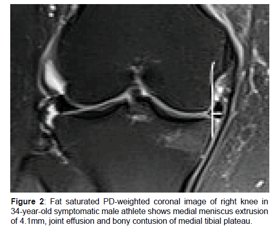
The results obtained from the 60 cases with knee injuries caused by sport (football playing ) demonstrated a significant association between meniscal extrusion and medial meniscus involvement [Figure 2], meniscal tear, medial meniscus tear, and types of meniscal tear (p<0.05). However, there was no significant association between medial meniscal extrusion, ACL tear, PCL tear, MCL tear, LCL tear, age, and joint effusion (p>0.05). Table 3 shows the statistical correlation of both subgroups with the associated knee injuries. Table 4 shows the distribution of the associated meniscus tear types.
| Trauma | Extrusion | Total | P-value | ||
|---|---|---|---|---|---|
| Yes | No | ||||
| Gender | Male | 12 (35.3) | 22 (64.7) | 34 (100.0) | 0.046 |
| Female | 17 (60.7) | 11 (39.3) | 28 (100.0) | ||
| Total | 29 (46.8) | 33 (53.2) | 62 (100.0) | ||
| Medial meniscus extrusion | Yes | 28 (100.0) | 0 (0.0) | 28 (100.0) | 0.001 |
| No | 1 (2.9) | 33 (97.1) | 34 (100.0) | ||
| Total | 29 (46.8) | 33 (53.2) | 62 (100.0) | ||
| Lateral meniscus extrusion | Yes | 1 (100.0) | 0 (0.0) | 1 (100.0) | 0.468 |
| No | 28 (45.9) | 33 (54.1) | 61 (100.0) | ||
| Total | 29 (46.8) | 33 (53.2) | 62 (100.0) | ||
| Meniscal tear | Yes | 27 (75.0) | 9 (25.0) | 36 (100.0) | 0.001 |
| No | 2 (7.7) | 24 (92.3) | 26 (100.0) | ||
| Total | 29 (46.8) | 33 (53.2) | 62 (100.0) | ||
| Medial meniscus tear | Yes | 26 (76.5) | 8 (23.5) | 34 (100.0) | 0.001 |
| No | 3 (10.7) | 25 (89.3) | 28 (100.0) | ||
| Total | 29 (46.8) | 33 (53.2) | 62 (100.0) | ||
| Lateral meniscus tear | Yes | 1 (50.0) | 1 (50.0) | 2 (100.0) | 1.000 |
| No | 28 (46.7) | 32 (53.3) | 60 (100.0) | ||
| Total | 29 (46.8) | 33 (53.2) | 62 (100.0) | ||
| Joint effusion | Yes | 21 (61.8) | 13 (38.2) | 34 (100.0) | 0.009 |
| No | 8 (28.6) | 20 (71.4) | 28 (100.0) | ||
| Total | 29 (46.8) | 33 (53.2) | 62 (100.0) | ||
| ACL tear | No | 18 (48.6) | 19 (51.4) | 37 (100.0) | 0.937 |
| High grade | 4 (44.4) | 5 (55.6) | 9 (100.0) | ||
| Low grade | 7 (43.8) | 9 (56.3) | 16 (100.0) | ||
| Total | 29 (46.8) | 33 (53.2) | 62 (100.0) | ||
| PCL tear | No | 22 (42.3) | 30 (57.7) | 52 (100.0) | 0.167 |
| Low grade | 7 (70.0) | 3 (30.0) | 10 (100.0) | ||
| Total | 29 (46.8) | 33 (53.2) | 62 (100.0) | ||
| MCL tear | Yes | 2 (100.0) | 0 (0.0) | 2 (100.0) | 0.215 |
| No | 27 (45.0) | 33 (55.0) | 60 (100.0) | ||
| Total | 29 (46.8) | 33 (53.2) | 62 (100.0) | ||
| LCL tear | Yes | 1 (100.0) | 0 (0.0) | 1 (100.0) | 0.468 |
| No | 28 (45.9) | 33 (54.1) | 61 (100.0) | ||
| Total | 29 (46.8) | 33 (53.2) | 62 (100.0) | ||
| Athletes | Extrusion | Total | P-value | ||
| Yes | No | ||||
| Medial meniscus extrusion | Yes | 8 (100.0) | 0 (0.0) | 8 (100.0) | 0.001 |
| No | 0 (0.0) | 52 (100.0) | 52 (100.0) | ||
| Total | 8 (13.3) | 52 (86.7) | 60 (100.0) | ||
| Meniscal tear | Yes | 8 (34.8) | 15 (65.2) | 23 (100.0) | 0.001 |
| No | 0 (0.0) | 37 (100.0) | 37 (100.0) | ||
| Total | 8 (13.3) | 52 (86.7) | 60 (100.0) | ||
| Medial meniscus tear | Yes | 8 (36.4) | 14 (63.6) | 22 (100.0) | 0.001 |
| No | 0 (0.0) | 38 (100.0) | 38 (100.0) | ||
| Total | 8 (13.3) | 52 (86.7) | 60 (100.0) | ||
| Lateral meniscus tear | Yes | 0 (0.0) | 1 (100.0) | 1 (100.0) | 1.000 |
| No | 8 (13.6) | 51 (86.4) | 59 (100.0) | ||
| Total | 8 (13.3) | 52 (86.7) | 60 (100.0) | ||
| Joint effusion | Yes | 6 (75.0) | 20 (38.5) | 26 (43.3) | 0.06* |
| No | 2 (25.0) | 32 (61.5) | 34 (56.7) | ||
| Total | 8 (100.0) | 52 (100.0) | 60 (100.0) | ||
| ACL tear | No | 3 (13.6) | 19 (86.4) | 22 (100.0) | 0.730 |
| High grade | 2 (9.1) | 20 (90.9) | 22 (100.0) | ||
| Low grade | 3 (18.8) | 13 (81.3) | 16 (100.0) | ||
| Total | 8 (13.3) | 52 (86.7) | 60 (100.0) | ||
| PCL tear | No | 8 (13.8) | 50 (86.2) | 58 (100.0) | 1.000 |
| High grade | 0 (0.0) | 1 (100.0) | 1 (100.0) | ||
| Low grade | 0 (0.0) | 1 (100.0) | 1 (100.0) | ||
| Total | 8 (13.3) | 52 (86.7) | 60 (100.0) | ||
| MCL tear | Yes | 1 (33.3) | 2 (66.7) | 3 (100.0) | *0.354 |
| No | 7 (12.3) | 50 (87.7) | 57 (100.0) | ||
| Total | 8 (13.3) | 52 (86.7) | 60 (100.0) | ||
Table 3: Correlation between subgroups and the associated knee injuries.
| Athletes | Extrusion | Total | P-value | ||
|---|---|---|---|---|---|
| Yes | No | ||||
| Type of meniscal tear | No | 0 (0.0) | 37 (71.2) | 37 (61.7) | 0.001 |
| BHT | 0 (0.0) | 3 (5.8) | 3 (5.0) | ||
| Complex tear of AH, BHT | 0 (0.0) | 1 (1.7) | 1 (1.7) | ||
| Complex tear of PH | 0 (0.0) | 3 (5.8) | 3 (5.0) | ||
| Complex tear of PH, BHT | 0 (0.0) | 1 (1.7) | 1 (1.7) | ||
| Horizontal tear of PH | 1 (12.5) | 4 (7.7) | 5 (8.3) | ||
| Radial tear | 2 (25.0) | 0 (0.0) | 2 (3.3) | ||
| Root tear | 4 (50.0) | 0 (0.0) | 4 (6.7) | ||
| Vertical tear of PH | 1 (12.5) | 3 (5.8) | 4 (6.7) | ||
| Total | 8 (100.0) | 52 (100.0) | 60 (100.0) | ||
| Trauma | Extrusion | Total | P-value | ||
| Yes | No | ||||
| Type of meniscal tear | No | 0 (0.0) | 24 (100.0) | 24 (100.0) | 0.001 |
| Complex tear of PH | 5 (62.5) | 3 (37.5) | 8 (100.0) | ||
| Horizontal tear of AH | 0 (0.0) | 1 (100.0) | 1 (100.0) | ||
| Horizontal tear of PH | 2 (40.0) | 3 (60.0) | 5 (100.0) | ||
| Intra-substance degeneration | 2 (100.0) | 0 (0.0) | 2 (100.0) | ||
| Radial tear | 5 (100.0) | 0 (0.0) | 5 (100.0) | ||
| Radial tear , BHT | 2 (100.0) | 0 (0.0) | 2 (100.0) | ||
| Root tear | 10 (100.0) | 0 (0.0) | 10 (100.0) | ||
| Root tear , BHT | 2 (100.0) | 0 (0.0) | 2 (100.0) | ||
| Vertical tear of PH | 1 (33.3) | 2 (66.7) | 3 (100.0) | ||
| Total | 29 (46.8) | 33 (53.2) | 62 (100.0) | ||
Table 4: The distribution of the associated meniscus tears types.
Discussion
The present study was an investigation into the prevalence of meniscal extrusion and associated knee joint abnormalities in patients with a history of knee trauma. The prevalence of meniscus extrusion in our overall study sample was 30.3%, athletes showed 13.3% prevalence versus 46.8% in non-athlete trauma group, while Rennie and Finlay (2006) reported 48% prevalence in athletes versus 30% in the control subjects. [18]
Most of the study sample were adults with an age range of 25-64 years. Similarly, Guermazi et al. reported a higher prevalence of knee trauma among adults compared to other age groups. [5] It was also seen that most of the patients (77%) were males. This finding is not in line with the reports by Shultz et al. who stated meniscal extrusion is more prevalent among women than men, due to the higher laxity of special knee structures like collateral ligaments in women compared to men. [14] This difference can be attributed to the fact that the present study was participated by a remarkably higher number of men than women.
It was observed that meniscus extrusion was significantly more common in the medial than the lateral meniscus and this finding is in good agreement with the study conducted by Puig et al. who reported medial and lateral extrusion respectively in 68.5% and 18.8% of the studied patients. [19]
Regarding the association between meniscus extrusion, meniscus tear and its type, a statistically significant association was noticed, and this is in line with several studies which referred to as tear of medial menisci as the most important risk factor for meniscal extrusion. [20-23] Root type tear was the most prevalent in our study group, followed by complex tear posterior horn of medial meniscus, and horizontal tear posterior horn of medial meniscus. Similar results were reported by other previously conducted studies. [24,25]
Our study showed a significant relationship between gender and meniscal extrusion. This finding is not in agreement with the one reported in the study conducted by Ding et al. who concluded that age, sex and family history of OA do not have a significant association with baseline medial meniscal extrusion. [26] This difference can be attributed to the fact that the present study was participated mostly by males (77%), while the number of females in the study carried out by Ding et al. as more than males (58% vs. 42%).
Moreover, there was a significant relationship between meniscal extrusion and types of meniscal tear. In this regard, meniscal root tears were found to be quite prevalent in cases with meniscal extrusion. Similarly, Carreau et al. reported that tear of the meniscus root can lead to meniscal extrusion. [27] Joint effusion was also significantly associated with meniscal extrusion. In addition, posterior cruciate ligament tear was found to be significantly associated with meniscal extrusion. In line with these two findings, Crema et al. showed a significant association between meniscal extrusions, joint effusion and posterior cruciate ligament tear. [15] A significant relationship was also observed between age and development of meniscal extrusion, such that meniscal extrusion was more prevalent among adults. Similar results were reported by Englund et al. [28] The results revealed that meniscal extrusion did not have a significant relationship with ACL, MCL, and LCL tears. This finding is supported by the study conducted by Hulet et al. [29]
Meniscus extrusion and trauma groups: analyzing the association between meniscal extrusion and the studied variables in the group who had knee trauma due to falling, similar significant association was found as for all of the 122 cases, except for the association between meniscal extrusion and posterior cruciate ligament tear (PCL), which was significant for the 122 cases (62 cases with knee injury from falling on ground and 60 from football playing). However, this association was not significant for the cases that had knee trauma as a result of falling on ground. Although this association was not significant, meniscal extrusion was seen in 42.3% of the cases without PCL tear. This finding is in line with the study carried out by Crema et al. [15]
Meniscus extrusion and trauma by football playing group: the results obtained for the cases with knee trauma caused by football playing indicated almost similar results as obtained for all of the 122 cases and the of knee injuries from falling on ground, except for the non-significant correlation of meniscal extrusion with posterior cruciate ligament tear, joint effusion, and age. Although the association between meniscal extrusion and joint effusion was not significant, 75% of the cases with joint effusion had experienced meniscal extrusion; our result was not in line with Rennie and Finlay who reported a significant association between meniscal extrusion and joint effusion in athletes. [18] The results also showed that the group with knee trauma caused by falling on ground and the group with knee injury as a result of football playing were significantly different in terms of their age and meniscal extrusion distance, such that the mean age of cases with knee injuries from falling on ground was 35 years, while that of the athletes was 28.43 years. Moreover, the meniscal extrusion distance was shorter in athletes compared to the other group, such that meniscal extrusion distance was 2.28 and 3.21 mm in the athletes and trauma group respectively. This finding is almost similar to the one reported by Rennie and Finlay. [18]
Conclusion
Prevalence of meniscal extrusion is high among cases with knee injuries whether due to falling on ground or football playing, medial meniscus involvement is much more commonly affected, with a significant association between meniscus extrusion and meniscus tear, root type tear, joint effusion and PCL tear. Medial meniscus tear is the most significant risk factors for meniscal excursion. Athletes experience a shorter meniscal extrusion distance compared to the non-athletes.
Competing Interests
The authors declare that they have no competing interests.
References
- Chawla A, Aggarwal AP. Menisci: Structure and Function. Asian Journal of Arthroscopy. 2016;1:3-7.
- Chivers MD, Howitt SD. Anatomy and physical examination of the knee menisci: a narrative review of the orthopedic literature. J Can Chiropr Assoc. 2009;53:319e33
- Ahmed AM. The load bearing role of the knee menisci. In: Mow VC, Arnoczky SP, Jackson DW, (eds). Knee meniscus: basic and clinical foundations. New York, NY: Raven Press, USA, 1992:59-73
- Bryceland JK, Powell AJ, Nunn T. Knee Menisci. Cartilage. 2017; 8:99-104.
- Guermazi A, Niu J, Hayashi D, Roemer FW, Englund M, Neogi T, et al. Prevalence of abnormalities in knees detected by MRI in adults without knee osteoarthritis: population based observational study (Framingham Osteoarthritis Study). BMJ. 2012;345:e5339.
- Englund M, Guermazi A, Gale D, Hunter DJ, Aliabadi P, Clancy M, et al. Incidental meniscal findings on knee MRI in middle-aged and elderly persons. N Engl J Med. 2008;359:1108e15
- Li TY. Distension of the medial collateral ligament bursa of the knee with possible bursitis: a sonographic case series. Journal of Diagnostic Medical Sonography. 2017;33:22-30.
- Muzaffar N, Kirmani O, Ahsan M, Ahmad S. Meniscal Extrusion in the Knee: Should only 3 mm Extrusion be Considered Significant? An Assessment by MRI and Arthroscopy. Malays Orthop J. 2015;9:17-20.
- Guermazi A, Hayashi D, Jarraya M, Roemer FW, Zhang Y, Niu J, et al. Medial posterior meniscal root tears are associated with development or worsening of medial tibiofemoral cartilage damage: the multicenter osteoarthritis study. Radiology. 2013;268:814e21.
- Allen DM, Li L, Crema MD, Marra MD, Guermazi A, Wyman BT, et al. The relationship between meniscal tears and meniscal position. Therapeutic advances in musculoskeletal disease. 2010;2:315-323.
- Englund M, Felson DT, Guermazi A, Roemer FW, Wang K, Crema MD, et al. Risk factors for medial meniscal pathology on knee MRI in older US adults: a multicenter prospective cohort study. Ann Rheum Dis. 2011;70:1733-1739
- Blöcker K, Guermazi A, Wirth W, Benichou O, Kwoh CK, Hunter DJ, et al. Tibial coverage, meniscus position, size and damage in knees discordant for joint space narrowing–data from the Osteoarthritis Initiative. Osteoarthritis and Cartilage. 2013;21:419-427.
- Bruns K, Svensson F, Turkiewicz A, Wirth W, Guermazi A, Eckstein F, et al. Meniscus body position and its change over four years in asymptomatic adults: a cohort study using data from the Osteoarthritis Initiative (OAI). BMC Musculoskelet Disord. 2014;15:32.
- Shultz SJ, Kirk SE, Johnson ML, Sander TC, Perrin DH. Relationship between sex hormones and anterior knee laxity across the menstrual cycle. Med Sci Sports Exerc. 2004;36:1165e74.
- Crema MD, Roemer FW, Felson DT, Englund M, Wang K, Jarraya M, et al. Factors associated with meniscal extrusion in knees with or at risk for osteoarthritis: the Multicenter Osteoarthritis study. Radiology. 2012;264:494-503.
- Roemer FW, Felson DT, Yang T, Niu J, Crema MD, Englund M, et al. The association between meniscal damage of the posterior horns and localized posterior synovitis detected on T1-weighted contrast-enhanced MRI—the MOST study. In: Seminars in arthritis and rheumatism 2013;42:573-581.
- Breitenseher MJ, Trattni S, Dobrocky I, Kukla C, Nehrer S, Steiner E, et al. MR imaging of meniscal subluxation in the knee. Acta Radiologica. 1997;38:876-879.
- Rennie WJ, Finlay DBL. Meniscal extrusion in young athletes: associated knee joint abnormalities. American Journal of Roentgenology. 2006;186:791-794.
- Puig L, Monllau JC, Corrales M, Pelfort X, Melendo E, Cáceres E. Factors affecting meniscal extrusion: correlation with MRI, clinical, and arthroscopic findings. Knee Surg Sports Traumatol Arthrosc. 2006;14:394-398.
- Lerer DB, Umans HR, Hu MX, Jones MH. The role of meniscal root pathology and radial meniscal tear in medial meniscal extrusion. Skeletal Radiol. 2004;33 (10):569–574
- Lee DH, Lee BS, Kim JM, Yang KS, Cha EJ, Park JH, et al. Predictors of degenerative medial meniscus extrusion: radial component and knee osteoarthritis. Knee Surgery, Sports Traumatology, Arthroscopy. 2011;19:222-229.
- Brody JM, Lin HM, Hulstyn MJ, Tung GA. Lateral meniscus root tear and meniscus extrusion with anterior cruciate ligament tear. Radiology. 2006;239:805-810
- Pache S, Aman ZS, Kennedy M, Nakama GY, Moatshe G, Ziegler C, et al. Meniscal root tears: current concepts review. Archives of Bone and Joint Surgery. 2018;6:250.
- Moatshe G, Chahla J, Slette E, Engebretsen L, Laprade RF. Posterior meniscal root injuries. Acta Orthop. 2016;87:452-458.
- Ahmed AF, Azeem AA, Eladawy A, Abdeen M. MRI as an accurate tool for the diagnosis and characterization of different knee joint meniscal injuries. The Egyptian Journal of Radiology and Nuclear Medicine. 2017;48:953-960.
- Ding C, Martel-Pelletier J, Pelletier JP, Abram F, Raynauld JP, Cicuttini F, et al. Knee meniscal extrusion in a largely non-osteoarthritic cohort: association with greater loss of cartilage volume. Arthritis Research & Therapy. 2007;9:R21.
- Carreau JH, Sitton SE, Bollier M. Medial meniscus root tear in the middle aged patient: a case based review. Iowa Orthop J. 2017;37:123-132.
- Englund M, Guermazi A, Gale D, Hunter DJ, Aliabadi P, Clancy M, et al. Incidental meniscal findings on knee MRI in middle-aged and elderly persons. N Engl J Med. 2008;359:1108-1115
- Hulet C, Pereira H, Peretti G, Denti M. Surgery of the Meniscus, ESSKA Book. 2016.


 The Annals of Medical and Health Sciences Research is a monthly multidisciplinary medical journal.