Triorchidism with Torsion
- *Corresponding Author:
- Dr. Kishore Kumar BN
76, 11th Cross, Indira Nagar I Stage, Bangalore-560 038, India.
E-mail: kishoreradiologist@gmail.com
Citation: Kumar K, Das D, S. Triorchidism with torsion.Ann Med Health Sci Res 2012;2:199-201.
Abstract
Polyorchidism is a rare anomaly; triorchidism is the commonest type and is frequently associated with cryptorchidism, inguinal hernia, and testicular torsion. It is also reported as an increased risk of testicular malignancy. We report a case of 72‑year‑old man with left supernumerary testes in the left hemiscrotum. He presented with pain in his left hemiscrotum of short duration. Physical examination revealed tender hemiscrotum. Both ultrasound and MRI examinations revealed polyorchidism. One of the testes in the left hemiscrotum revealed features of torsion and hemorrhagic necrosis. Polyorchidism is a rare entity. In most cases, sonography alone is diagnostic of polyorchidism. Magnetic resonance imaging may provide additional information in complicated cases of polyorchidism
https://linktr.ee/marmaris1 https://www.divephotoguide.com/user/marmaris1/ https://artmight.com/user/profile/2408931 https://allmyfaves.com/marmaris1 https://www.fimfiction.net/user/628252/marmaris https://www.drupalgovcon.org/user/568666 https://www.roleplaygateway.com/member/marmaris1/ https://www.kickstarter.com/profile/825997479/about https://tapas.io/sam659667 https://seedandspark.com/user/marmaris1 https://starity.hu/profil/386591-marmaris1/ https://www.informationweek.com/profile.asp https://nootheme.com/forums/users/marmaris1/ https://app.roll20.net/users/12308972/marmaris1-m https://www.360cities.net/profile/sam659667 https://fileforum.com/profile/marmaris1 https://wordpress.org/support/users/marmaris1/ https://www.hebergementweb.org/members/marmaris1.539143/ https://forum.cs-cart.com/u/marmaris1/ https://www.tntxtruck.com/User-Profile/UserId/12208 https://calis.delfi.lv/blogs/posts/205779-linkler/lietotajs/310821-marmaris1/ https://profile.ameba.jp/ameba/marmaris1/ https://www.avianwaves.com/User-Profile/userId/184770 https://engine.eatsleepride.com/rider/marmaris1 https://keymander.iogear.com/profile/53051/marmaris1 https://www.mifare.net/support/forum/users/marmaris1/ https://inkbunny.net/marmaris1?&success=Profile+settings+saved. https://www.diggerslist.com/64e0c4fad170b/about https://research.openhumans.org/member/me/ http://bluerevolutioncrowdfunding.crowdfundhq.com/users/marmaris1 https://www.cakeresume.com/me/marmaris1 https://educatorpages.com/site/marrmaris1/pages/about-me? https://gotartwork.com/Profile/marmaris1-marmaris1/253385/ https://www.facer.io/user/OGfo7VWrhR https://able2know.org/user/marmaris1/ https://www.techrum.vn/members/marmaris1.230749/#about http://foxsheets.statfoxsports.com/UserProfile/tabid/57/userId/145739/Default.aspx http://riosabeloco.com/User-Profile/userId/196075 http://phillipsservices.net/UserProfile/tabid/43/userId/245691/Default.aspx http://www.ramsa.ma/UserProfile/tabid/42/userId/962725/Default.aspx http://krachelart.com/UserProfile/tabid/43/userId/1242308/Default.aspx http://kedcorp.org/UserProfile/tabid/42/userId/72907/Default.aspx http://atlantabackflowtesting.com/UserProfile/tabid/43/userId/561626/Default.aspx https://www.intensedebate.com/people/marmaris12 https://rosalind.info/users/marmaris1/ https://wordpress.com/me https://photozou.jp/user/top/3342211
Keywords
Magnetic resonance imaging, Polyorchidism, Sonography, Torsion, Triorchidism
Introduction
Polyorchidism, or supernumerary testes is a rare condition, which is embryological in origin. It results from an abnormal division of the genital ridge. Although three testes is the most common form, as many as five have been reported. In approximately 75% of cases, the supernumerary testes are intrascrotal, and the patients most often present with a painless scrotal mass. Of the remaining cases, 20% of the testes are inguinal and 5% are retroperitoneal.[1,2] Supernumerary testes are retractile and more mobility is at increased risk for torsion. An increased prevalence of carcinoma has also been reported.[1]
Case Report
A 72‑year‑old man presented with vague pain in the lower abdomen and scrotum. There was no history of trauma and he stated that the pain was severe in nature and had been present for a short time. He was married and had three children. Physical examination revealed swelling of scrotum on the left side. There were no features to suggest inguinal hernia. He was referred to the radiology department for evaluation of pain.
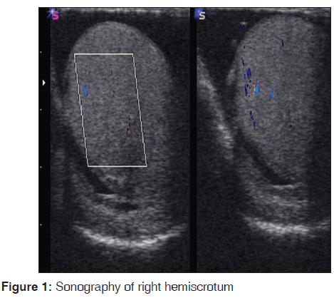
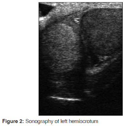
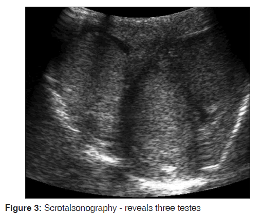
Ultrasonography of abdomen was normal. Gray scale and color Doppler sonography of right hemiscrotum revealed a normal testicle with homogenous texture, measuring about 3.8 × 3 cm. Normal intratesticular flow was noted. Epididymis was normal in size and echotexture. Minimal hydrocele was noted [Figure 1]. Left hemiscrotum revealed two testicles The lower testis showed homogenous echotexture, measuring 3.7 × 3 cm, with normal intratesticular flow. The upper testis measured 3.2 × 2.4 cm and showed homogenous hypoechogenicity with no detectable intratesticular flow compatible with ischemia [Figures 2 and 3]. Epididymis was single and appeared normal. Minimal hydrocele was noted.
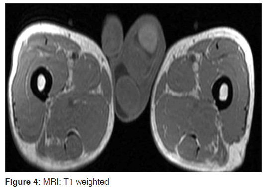
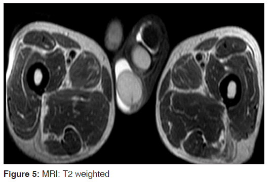
Magnetic resonance imaging (MRI) examination was done, which showed normal T 1 and T 2 images of right testis and lower testis of left hemiscrotum [Figure 4]. The upper testis of left hemiscrotum showed increase signal intensity on T1 and decreased signal intensity on T2 images, suggestive of torsion and hemorrhagic necrosis [Figure 5]. Minimal hydrocele was noted in both hemiscrota.
Discussion
On the basis of embryologic development, Leung classified polyorchidism into four types [3] In type A, the division separates a small part of the genital ridge, which does not contact the mesonephric duct. Therefore, the supernumerary testis lacks an epididymis and vas deferens. In type B, the division of the genital ridge occurs in the region where the primordial gonads are attached to the mesonephric ducts and the supernumerary testis has its own epididymis. In type C, the supernumerary testis has its own epididymis and shares the vas deferens with the regular testis in a parallel fashion. In this type of polyorchidism, there is an incomplete longitudinal division of the genital ridge and the proximal portion of the mesonephric duct. In type D, which is the least common, complete longitudinal duplication of the genital ridge and mesonephric duct occurs, with resultant complete duplication of testes, epididymides, and vas deferens. Further investigation could not be done in our case to relate as per Leung classification.
Triorchidism is the most common type of polyorchidism and presents with two testes on one side (usually the left) and one testis on the other side. Rare case of polyorchidism with three homolateral testes on the right side and absent testis on the left side has been reported.[4]Splenogonadal fusion is a rare congenital anomaly that may sonographically resemble polyorchidism.[5,6] In this entity spleen, gonad, epididymis, and vas deferens are fused. Sonography reveals a mass with the testicle of similar echogenicity and it may mimic the appearance of polyorchidism. When splenogonadal fusion is suspected, a technetium sulfur colloid scan should be performed to confirm the presence of ectopic splenic tissue [5] Singer et al.[7] proposed a classification based on reproductive potential of the supernumerary testis. In type 1, the supernumerary testis has reproductive potential because of attachment to a draining epididymis and vas deferens. In type 2, the supernumerary testis has no reproductive potential because of lack of a draining system. Singer et al. further subclassified these types into A, if the supernumerary testis is located in the scrotum, or B, if it is ectopic The sonographic appearance of polyorchidism is presence of scrotal mass that has an echo pattern identical to that of the ipsilateral testicle.[8] MRI appearance is a round or oval shaped structure showing typical signal characteristics of testicles, i.e., homogeneous intermediate signal intensity on T1 weighted and high signal intensity on T2 weighted images.[9]
Polyorchidism is a rare genitourinary abnormality, and torsion is one of its associated complications; however, the diagnosis of polyorchidism with or without torsion can be made readily with ultrasonography when one is aware of this entity.[10]
Sonographic features of torsion are homogenous hypoechogenicity of testis with absent flow on color Doppler study. MRI features of torsion are increased signal intensity on T1 and decreased signal intensity on T2 weighted sequences.[11] Our case showed features of polyorchidism with torsion of one of the testes in left hemiscrotum, both on sonography and MRI.
Conclusion
Polyorchidism is a rare congenital anomaly. Torsion is one of the complications of this entity. Polyorchidism can be diagnosed on sonography. Torsion can be suspected and diagnosed on color Doppler study. MRI can be used for confirmation but may be more helpful in cases complicated by torsion, cryptorchidism, or neoplasia.
Source of Support: Nil.
Conflict of Interest: None declared.
References
- Ulbright TM, Amin MB, Young RH. Miscellaneous primary tumors of the testis, adnexa and spermatic cord. In: Rosai J,Sobin LH, editors. Atlas of tumor pathology, fasc 25, ser 3.Washington, DC: Armed Forces Institute of Pathology; 1999.p. 235-366.
- Bostwick DG. Spermatic cord and testicular adnexa. In:Bostwick DG, Eble JN, editors. Urologic surgical pathology.St Louis, Mo: Mosby; 1997. p. 647-74.
- Leung AK. Polyorchidism. Am Fam Physician 1988;38:153-6.4.
- Vijayanadh O, Shanbhogue AK, Doherty GP. An unusualcase of polyorchidism with three homolateral testes and contralateral anorchia. Eur J Radiol Extra 2009;72:e129-31.
- Akbar S, Sayyed T, Jafri S, Hasteh F, Neill JS. Multimodality imaging of paratesticular neoplasms and their rare mimics. Radiographics 2003;23:1461-76.
- Cirillo RL Jr, Coley BD, Binkovitz LA, Jayanthi RV. Sonographic findings in splenogonadal fusion. Pediatr Radiol 1999;29:73-5.
- Singer BR, Donaldson JG, Jackson DS. Polyorchidism: functional classification and management strategy. Urology 1992;39:384-8.
- Chung TJ, Yao WJ. Sonographic features of polyorchidism. J Clin Ultrasound 2002;30:106-8.
- Yal�inkaya S, Sahin C, Sahin AF. Polyorchidism: sonographic and magnetic resonance imaging findings. Can Urol Assoc J 2011;5:E84-6.
- Hwang S, Aronoff DR, Leonidas JC. Case 82: polyorchidism with torsion. Radiology 2005;235:433-5.
- Watanabe Y, Nagayama M, Okumura A, Amoh Y, Suga T, Terai A, et al. Imaging of testicular torsion: Features of testicular hemorrhagic necrosis and clinical outcomes. J Magn Reson Imaging 2007;26:100-8.



 The Annals of Medical and Health Sciences Research is a monthly multidisciplinary medical journal.