
Working Length Determination of Root Canal of Young Permanent Tooth: An In Vitro Study
- *Corresponding Author:
- Dr. Amish Diwanji
Department of Pedodontics and Preventive Dentistry, Faculty of Dental Science, Nadiad, Gujarat, India.
E-mail: amishdiwanji@yahoo.co.in
Abstract
Background: Determination of correct working length is one of the keys to success in endodontic therapy. Aim: The aim of this study was to evaluate the diagnostic efficacy of various methods to determine working length of root canal. Materials and Methods: Tactile method was assessed using digital radiography and compared with electronic method using apex locator. A total sample of 30 single rooted young permanent teeth the (mandibular first premolars) with matured apices were selected for the study. Access cavity preparation was carried out. Working length was measured by tactile method using digital radiography and electronic method using apex locator with no 15 K file. Actual working length was established by grinding of cementum and dentine from the root apex and was observed under stereomicroscope. Data was collected and statistical analysis was carried out with the help of SPSS‑15. Results: The results of this study showed that there was a significant difference between tactile method assessed by digital radiography and electronic method using apex locator. Conclusion: Apex locator was found to be more reliable and accurate when compared with the actual length.
Keywords
Apex locator, Digital radiography, Stereomicroscope, Working length determination
Introduction
An accurate working length determination of root canal during endodontic treatment is very essential. It makes endodontic treatment easier for an operator to remove necrotic tissue and prepare canals precisely.[1]
Various methods have been used to establish correct working length. These include use of conventional or digital radiography,[2] tactile method [3] and moisture on paper point.[4] All of these methods have their limitations. Radiographs are subjected to distortion and magnification. It comprises accurate location of root apices.[4] They are technique sensitive in exposure and interpretation. They also provide a two dimensional image of a three dimensional structure which may not represent real position of apical region. Furthermore, in many cases with conventional radiography, it is difficult to establish the actual length of the canal with a two dimensional image.[2,3] It becomes even more difficult to establish correct working length with radiography when root canal system is superimposed radiographically by anatomic structures. In such cases, electronic method using apex locator is very useful.[6]
With advancement in technology, a new method of establishing working length with electronic apex locater was introduced by Sunada in 1962. Since then, different generation of electronic apex locators have been developed to measure root canal length with superior accuracy.[7] Electronic apex locators give reliable results when apical foramen does not coincide with the anatomic root apex.
The cemento-dentinal junction (CDJ), where the pulp and periodontal tissue meet is considered as physiological limit for working length, biomechanical preparation and obturation in endodontic treatment. This landmark cannot be precisely determined radiographically [11] and has been claimed to be determined by modern electronic apex locators with more than 90% accuracy.[12] The location of apical constriction varies from root to root, hence its relationship with CDJ is also different as CDJ could be irregular and at different height on one wall of root compare with the opposite wall.[13,14]
Estimated working length can be determined with radiography and an electronic method, but correct working length can be determined only by comparing these two methods with direct visualization method (DVM) which can be controlled histologically under microscope. Exact position of file tip can be determined only when teeth are histologically examined after extraction.
Therefore, the study was aimed to compare exact working length of root canal with radiographic method and electronic method using apex locator and comparing both the methods with the actual length from histological sections under stereomicroscope.
Materials and Methods
A total number of 30 single rooted young permanent teeth (mandibular first premolars) with matured apices (age group 15-17 years), extracted for orthodontic purpose were taken. The study received the necessary ethical clearance from IRB committee of Darshan Dental College, Udaipur, India. Teeth were cleaned from any residual tissues and cleaned with water and stored in 10% formalin solution. Conventional access cavity preparation was carried out on all teeth using diamond burs, high speed hand piece and water coolant. The cusps of teeth were flattened using a high speed hand piece to establish a surface level to serve as a stable and equal platform for all measurements. Pulp chamber wall was smoothened and root canal orifices were identified. Canals were irrigated with 2.5% sodium hypochlorite. Pulp tissues were extirpated using barbed broaches without making any attempt to enlarge the canals with endodontic instruments. All the samples were stabilized in an alginate mold and measurements were made within 2-3 h of the model being prepared. Tactile method was assessed by digital radiography and an electronic method was determined using apex locator (Propex™, Dentsply Maillefer).
Length determination by tactile method using digital radiography
Estimated working length was determined on the diagnostic radiograph. Canals were negotiated with no. 15 K file up to the estimated working length. File was pushed apically until the resistance was felt and canal patency was evaluated. Reference point was noted down. A good quality radiograph was taken by using digital radiography (RVG-satelec). Length was measured using Maillefer gauge (Endo block).
Length determination by apex locator
The same sample was evaluated for length determination by electronic method. The labial clip was fixed with the edge of saline bath in contact with fluid all the time. The apex locator was turned on and no. 15 K file was advanced into the root canal just beyond the foramen. As the instrument moved apically, the digital display indicated the distance from the tip of the file to the apical constriction in the tenth of a millimeter. The unit’s cable was clipped in to metal shank after removing 15 no K file.
The apex locator gave out signal in 3 ways, a digital display reading - 0, a pulsing audition, a flashing light. When the locator signaled the apical region; the rubber stop was adjusted at the same reference point. Length was measured and recorded. 1 mm was subtracted from that and the file was stabilized in canal using cyanoacrylate material within the chamber opening. The same procedure was followed for the rest of the samples.
Determination of actual length
The actual length was determined by selective grinding of tooth. After using electronic method, sample with stabilized file in a canal was carefully sectioned in a longitudinal direction against grinder and selective grinding was carried out until the canal was visible in the longitudinal direction. With diamond bur in straight hand piece, thin layer of dentin was removed and care was taken not to damage the instrument. Remaining thin layer of dentin was removed with probe and the canal was exposed with endodontic file within canal to observe the topography and relation between the file tip, apical constriction and anatomical apex under stereomicroscope [Figure 1]. The measurements were considered valid if the instrument remained stable for at least 5 s; otherwise value was recorded as an unstable measurement due to inability of apex locator to reveal a constant reading.

Figure 1: Determination of actual length and samples after selective grinding
Stereomicroscope observation
Samples were observed under stereomicroscope for the actual length determination [Figure 2]. The silicone stop was stabilized and distance between file tip, apical constriction, major foramen and the anatomical apex was measured under × 4.5 magnifications with a millimeter ruler to the nearest 0.25 mm. The data was statistically compared by ANOVA and paired t-test.

Figure 2: Stereomicroscopic observation
Data were collected and analyzed with SPSS-15.
Results
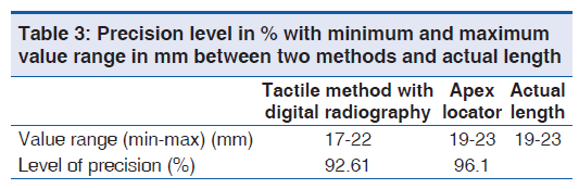
Table 1 represents the mean value and standard deviation of individual method. Mean value of apex locator was closer to the actual length as compared to tactile method confirmed by digital radiography. Comparison between tactile method and actual length was done and difference in the mean value was 1.45 and t value was −3.704, which was statistically significant. Similarly, comparison between apex locator and actual length done which showed a difference in the mean value was 0.40 and t value was −1.34 which was statistically non-significant. Table 2 summarizes one way analysis of variance for both methods included in the present study. ANOVA test showed F value of 7.44 which was highly significant statistically. Table 3 represents a range of minimum and maximum value among various methods and level of accuracy between them. Apex locator showed higher accuracy with 96.1%. Table 4 represents mean distance between file tip and actual length observed by radiographic method and electronic method using apex locator. Mean distance in radiographic method was 1.45 mm which was more than mean distance observed by electronic method (0.40 mm).
| Tactile method assessed by digital radiography | Apex locator | Actual length as seen on histological section | |
|---|---|---|---|
| Mean value (mm) | 19.90 | 20.95 | 21.35 |
| Standard deviation (mm) | 1.21 | 1.11 | 1.19 |
t value for tactile method assessed by digital radiography versus/actual length: 3.704. t value for apex locator versus/Actual length: 1.347
Table 1: Mean values of working length assessed by different methods
| Source of variation | Degree of freedom | Sum of square | Mean square | Standard deviation | P value |
|---|---|---|---|---|---|
| Between groups | 2 | 20.45 | 10.22 | <0.001 | |
| Within groups | 97 | 119.55 | 1.37 | 1.25 | |
| Total | 89 | 140 |
Table 2: Analysis of variance between two methods and within the method
| Distance between file tip and actual length assessed by tactile method (digital radiography apex) | Distance between file tip and actual length assessed by apex locator | |
|---|---|---|
| Mean (mm) | 1.45 | 0.40 |
| Standard | 0.98 | 0.31 |
| deviation (mm) |
Table 4: Comparison of mean distance in mm between file tip and apical foramen compared with the actual length in both techniques
Discussion
For successful endodontic treatment, establishing a correct working length is an important factor. Various studies have been done to evaluate the accuracy of working length of root canal during endodontic treatment. Apical constriction is an ideal spot for working length determination. It is a narrowest spot of root canal having lowest diameter of blood vessels, also known as minor diameter of the canal. It is proved that distance between apical constriction and external foramen is 0.5 to 1 mm [10]
K file (15 no.) was used as an endodontic instrument to determine working length. M A Martínez-Lozano et al.,[16] conducted a pilot study using different endodontic files to determine which file shows the most precise measurements. They recommended 15 no K file for working length determination in single rooted teeth. Further, it has been reported that electronic working length is not influenced by the size of measuring file used.[17]
The use of apex locator has gained a lot of attention while determining working length of canals during endodontic treatment.[18] In the present study, Propex (Dentsply Maillefer, Switzerland) apex locator was used, which is a multi-frequency based apex locator to determine root canal length. The calculation is based on the energy of the signal where the other apex locators usually use the amplitude signal. The manufacturer claims that energy measurement is more precise.
However, to establish the actual length with respect to CDJ, histological method has been recommended.[19] In the present study, samples were sectioned for histological approach in order to compare with the actual length. Martínez-Lozano et al.,[16] and Muthu et al.,[20] stated that histological method is the best approach to establish actual working length, i.e., by removing cementum and dentin. Apical foramen was considered to be standardized reference.
The samples were stabilized in alginate mold to prevent fluid movement inside the canal. It could be an important factor for registration of premature electronic reading using apex locator. This way an effort was made to overcomes the limitations of in vitro conditions. Measurements were made within 2-3 hr of the model being prepared to ensure the alginate was kept sufficiently humid.[23,24]
In the present study, distance between file tip and apical foramen was measured by radiographic method and an electronic method and compared them with the actual length. Apex locator showed higher accuracy with 96.1% as compared to digital radiography (92.6%). These results are comparable with that of Frank and Torabinejad [25] and Shabahang et al.,[26] who reported higher values in the range of 85% and 98% respectively.
As mentioned earlier, apical constriction is an ideal spot for working length in endodontic treatment which is 0.5 to 1 mm away from major foramen.[10-15] It is stated that apex locator can locate major foramen and a point between the apical constriction and the foramen depending on the resistance of the dentin.[27,28] Plotino et al.,[29] located apical constriction using Propex apex locator 0.5 mm short of the apex.
Recently, it has been recommended that canal preparation should be confined 1 mm short of electronic working length to avoid over preparation of apical region.[30] In the present study, major foramen was located by EAL and then 1 mm was subtracted in order to determine precise working length.
Conclusion
Within the limitations of this study, apex locator was found to be more reliable and accurate within 1mm of apical constriction when compared with radiographic method. However, working length determination should be carried out using a combination of both techniques. Further studies are still necessary.
Source of Support: Nil.
Conflict of Interest: None declared.
References
- Goldberg F, Marroquín BB, Frajlich S, Dreyer C. In vitro evaluation of the ability of three apex locators to determine the working length during retreatment. J Endod 2005;31:676-8.
- Stein TJ, Corcoran JF. Radiographic “working length” revisited. Oral Surg Oral Med Oral Pathol 1992;74:796-800.
- Seidberg BH, Alibrandi BV, Fine H, Logue B. Clinical investigation of measuring working lengths of root canals with an electronic device and with digital-tactile sense. J Am Dent Assoc 1975;90:379-87.
- Ruddle CJ. Cleaning and shaping root canal systems. In: Cohen S, Burns RC, editors. Pathways of the Pulp. 8th ed. St. Louis, MO: Mosby; 2002. p. 231-91.
- Kim E, Marmo M, Lee CY, Oh NS, Kim IK. An in vivo comparison of working length determination by only root-ZX apex locator versus combining root-ZX apex locator with radiographs using a new impression technique. Oral Surg Oral Med Oral Pathol Oral Radiol Endod 2008;105:e79-83.
- Custer LE. Exact method of locating apical foramen. J Natl Dent Assoc 1918;65:815-9.
- Sunada I. New method for measuring the length of the root canal. J Dent Res 1962;41:375-87.
- Gordon MP, Chandler NP. Electronic apex locators. Int Endod J 2004;37:425-37.
- Green D. A stereomicroscopic study of the root apices of 400 maxillary and mandibular anterior teeth. Oral Surg Oral Med Oral Pathol 1956;9:1224-32.
- Green D. Stereomicroscopic study of 700 root apices of maxillary and mandibular posterior teeth. Oral Surg Oral Med Oral Pathol 1960;13:728-33.
- Ricucci D, Langeland K. Apical limit of root canal instrumentation and obturation, part 2. A histological study. Int Endod J 1998;31:394-409.
- Bernardes RA, Duarte MA, Vasconcelos BC, Moraes IG, Bernardineli N, Garcia RB, et al. Evaluation of precision of length determination with 3 electronic apex locators: Root ZX, elements diagnostic unit and apex locator, and RomiAPEX D-30. Oral Surg Oral Med Oral Pathol Oral Radiol Endod 2007;104:e91-4.
- Dummer PM, McGinn JH, Rees DG. The position and topography of the apical canal constriction and apical foramen. Int Endod J 1984;17:192-8.
- Gutierrez JH, Aguayo P. Apical foraminal openings in human teeth. Number and location. Oral Surg Oral Med Oral Pathol Oral Radiol Endod 1995;79:769-77.
- Kuttler Y. Microscopic investigation of root apexes. J Am Dent Assoc 1955;50:544-52.
- Martínez-Lozano MA, Forner-Navarro L, Sánchez-Cortés JL, Llena-Puy C. Methodological considerations in the determination of working length. Int Endod J 2001;34:371-6.
- Nguyen HQ, Kaufman AY, Komorowski RC, Friedman S. Electronic length measurement using small and large files in enlarged canals. Int Endod J 1996;29:359-64.
- Kim E, Lee SJ. Electronic apex locator. Dent Clin North Am 2004;48:35-54.
- Hoer D, Attin T. The accuracy of electronic working length determination. Int Endod J 2004;37:125-31.
- Muthu S, Rajendran N, Rajan M, Sundaresan B. Evaluation of working length determination methods: An in vivo/ex vivo study. J Indian Soc Pedod Prev Dent 2007;18:60-2.
- Mayeda DL, Simon JH, Aimar DF, Finley K. In vivo measurement accuracy in vital and necrotic canals with the Endex apex locator. J Endod 1993;19:545-8.
- Pratten DH, McDonald NJ. Comparison of radiographic and electronic working lengths. J Endod 1996;22:173-6.
- Lucena-Martín C, Robles-Gijón V, Ferrer-Luque CM, de Mondelo JM. In vitro evaluation of the accuracy of three electronic apex locators. J Endod 2004;30:231-3.
- Tinaz AC, Alaçam T, Topuz O. A simple model to demonstrate the electronic apex locator. Int Endod J 2002;35:940-5.
- Frank AL, Torabinejad M. An in vivo evaluation of Endex electronic apex locator. J Endod 1993;19:177-9.
- Shabahang S, Goon WW, Gluxkin AH. An in vivo apex locator Root ZX. J Endod 1997;15:35-40.
- Lee SJ, Nam KC, Kim YJ, Kim DW. Clinical accuracy of a new apex locator with an automatic compensation circuit. J Endod 2002;28:706-9.
- Ounsi HF, Naaman A. In vitro evaluation of the reliability of the Root ZX electronic apex locator. Int Endod J 1999;32:120-3.
- Plotino G, Grande NM, Brigante L, Lesti B, Somma F. Ex vivo accuracy of three electronic apex locators: Root ZX, elements diagnostic unit and apex locator and propex. Int Endod J 2006;39:408-14.
- Guise GM, Goodell GG, Imamura GM. In vitro comparison of three electronic apex locators. J Endod 2010;36:279-81.


 The Annals of Medical and Health Sciences Research is a monthly multidisciplinary medical journal.