
Normal Range of Inferior Facial Angle (IFA) During the First Trimester to the Diagnosis of Micrognathia
Received: 01-Apr-2022, Manuscript No. amhsr-22-52341; Editor assigned: 04-Apr-2022, Pre QC No. amhsr-22-52341(PQ); Accepted Date: Apr 29, 2022 ; Reviewed: 19-Apr-2022 QC No. amhsr-22-52341; Revised: 25-Apr-2022, Manuscript No. amhsr-22-52341(R); Published: 29-Apr-2022, DOI: 10.54608.annalsmedical.2022.36
Citation: Asl LY, et al. Normal Range of Inferior Facial Angle (IFA) During the First Trimester to the Diagnosis of Micrognathia. Ann Med Health Sci Res. 2022;12:121-124.
This open-access article is distributed under the terms of the Creative Commons Attribution Non-Commercial License (CC BY-NC) (http://creativecommons.org/licenses/by-nc/4.0/), which permits reuse, distribution and reproduction of the article, provided that the original work is properly cited and the reuse is restricted to noncommercial purposes. For commercial reuse, contact reprints@pulsus.com
Abstract
Introduction: Fetal facial malformations such as micrognathia, maxillary dysplasia, cleft lip and palate, as well as absence of nasal bone have been defined to be associated with some chromosomal abnormalities or genetic syndrome. Ultrasonography can show these parameters exactly. Proper imaging allows medical staff and radiologists to better examine the anatomy of the fetal face and make a more accurate diagnosis. The current study aimed at establishing the reference range for IFA and analyzing its association with fetal micrognathia during the first trimester.
Materials & Methods: This cross-sectional study was performed on pregnant women with a gestational age of 18 to 28 weeks referred to Shahid Akbarabadi Hospital in Tehran. After visiting, informations including age of mother and gestational age was recorded. Then results of ultrasonography test was registered. After childbirth, infants were examed and assigned in to one of the 2 groups. After that, records of IFA were analyzed in both groups.
Results: In the present study, 329 eligible mothers included in the study. We recorded their information and IFA of their fetus. Average age of mothers was determined 30/996/20. Furthermore, gestational age ranged from 17 w+3 d to 27 w. Moreovere, minimum and maximum of IFA were 48 and 79, respectively. Average of IFA was also recorded to be 60/865/69. 2 of the fetuses had abnormality after birth (IFAs 48 and 49). This study showed a significant association of IFA with abnormality of mandible after birth and abnormality of mandible in fetus with lower IFA was found to be more probable.
Conclusion: Our results showed an average of 60/865/69 for IFA, indicating that average of IFA in Iranian fetuses is less than most of the world. This study also revealed a significant association of IFA with abnormality of mandible after birth, Howevere, comprehensive studies with large abnormal samples are nedeed to assess fetuses with lower IFA.
Keywords
Mandible; Ultrasonography; Gestational age; Inferior facial angle
Introduction
The development of the mandible occurs in the sixth gestational week and this complex developmental cascade depends on the fusion of various different embryonic components and complex interactions involving ectoderm of branchial arches and neural crest cells originating in the dorsal neural tube. [1,2]
As a matter of fact, several elements are involved in fetal mandible formation from different embryonic components to interact and fuse, where is more likely to be susceptible to a series of molecular and genetic insults. [1] Mandibular changes should be considered as alarm bells because they are associated with many syndromes and abnormalities. Jaw and facial abnormalities have a direct impact on fetal survival and can lead to numerous respiratory and swallowing problems. Fetal facial malformations such as micrognathia, maxillary dysplasia, cleft lip and palate, as well as absence of nasal bone have been defined to be associated with some chromosomal abnormalities or genetic syndrome. [3,4]
One of these abnormalities, including micrognatia, is a subtle facial abnormality featured by a small mandible and a receding chin. The micrognathia that occurs following neural crest cell hypoplasia can occur independently but is often associated with other abnormalities, e.g., chromosomal abnormalities, skeletal dysplasia, and various syndromes (Pierre Robin sequence, trisomy 13 and 18). [5-8]
Nowadays, micrognathia can be diagnosed via ultrasonography because of its typical ultrasonography features. [2] The Inferior Facial Angle (IFA), measured in a sagittal view of the facial profile, is capable of identifying micrognathia, leading to the differential diagnosis. [8] The jaw index, an axial view of the fetal mandible and the mandibular/maxillar width, can be helpful in assessing the severity of the micrognathia. Fetal Magnetic Resonance Imaging (MRI) can be helpful to assess the severity of fetal micrognathia evaluating a potential obstruction of the airway. [9] Proper imaging allows medical staff and radiologists to better examine the anatomy of the fetal face and make a more accurate diagnosis. [10] Nonetheless, prenatal diagnosis of fetal facial abnormalities in the first trimester is still a challenge, and a number of reliable and repeatable objective parameters are not accessible. In this study, fetal facial marker including IFA were measured in fetal facial mid-sagittal section [Figure 1].

Figure 1: Linear regression between the two variables gestational age and IFA in the subjects.
The current study aimed at establishing the reference range for IFA and analyzing its association with fetal micrognathia during the first trimester. As a matter of fact, the present study was conducted to find the normal range of IFA in Iranian embryos and to provide a suitable diagnostic test.
Materials and Methods
Sampling
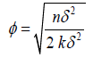
This cross-sectional study was performed on pregnant women with a gestational age of 18 weeks to 28 weeks referred to Shahid Akbarabadi Hospital in Tehran. According to the studies performed, the sample size of 300 was determined using the following formula:
The required sample is also collected by convenience sampling method. The checklists are then completed and the tests performed.
Data collection
After obtaining permission from the research assistant and relevant authorities, the information of patients referred to the relevant ward in Shahid Akbarabadi Hospital was extracted and the IFA was measured in normal fetuses.
Inclusion criteria included mothers with a gestational age of 18 weeks to 28 weeks and normal screening ultrasound. Exclusion criteria include: embryos with abnormalities
After birth, the infants examined and placed in two groups of abnormal and healthy. After birth, infants are examined for micrognathia and swallowing to compare the normal range of the IFA for both healthy and abnormal groups. The necessary information such as age and gestational age were recorded. Then the sonographic test was performed and the results were recorded in a checklist.
IFA was measured during the first trimester when the fetus was examined for nuchal translucency and nasal bone. The midsagittal surface was precisely defined by the baby tip in the front, the translucent diencephalon in the middle, the nuchal membrane in the back, and the roof of the mouth.
IFA is measured by defining a right-angled line from the vector drawn at the synostosis of the nasal bones to the vertical part of the forehead, at the synostosis level of the nasal bones, and the vector joining the tip of the mentum the anterior margin of the more protruding part of the lip.
Then the normal range of IFA and its condition in the patient group was examined. Naturally, an appropriate test would be one that identifies more patients and is positive in a smaller number of healthy people.
Data analysis
The information received in the answer sheet and test results were entered into SPSS software version 21. Then, the relationship between the desired factors is statistically examined by Chisquare test. All tests are performed by considering a significance level alpha=0.05.
Ethical considerations
Patients 'personal information was kept throughout the research and the individuals' information was entered into the software with a checklist code.
Results
In this study, we examined 328 people who were eligible to participate in the study. Preliminary data were recorded and fetal IFA was measured. The mean age of mothers was found to be 30.99 ± 6.20. The gestational age of the subjects was from 17 w+3 d to 27 w. Tables 1 and 2 show the demographic information of the subjects.
| Table 1: Age of mothers participating in the study. | |||
|---|---|---|---|
| Variable | Min | Max | Mean |
| Age of mothers | 16 | 46 | 30.99 ± 6.2 |
| Table 2: Gestational age of mothers participating in the study. | ||
|---|---|---|
| Gestational age | Number | Percent |
| 17w+3d | 1 | 0.3 |
| 17w+4d | 2 | 0.6 |
| 17w+5d | 5 | 1.5 |
| 17w+6d | 4 | 1.2 |
| 18w | 26 | 70.9 |
| 18w+1d | 16 | 40.9 |
| 18w+2d | 26 | 70.9 |
| 18w+3d | 31 | 9.4 |
| 18w+4d | 22 | 6.7 |
| 18w+5d | 28 | 8.5 |
| 18w+6d | 23 | 7 |
| 19w | 32 | 9.7 |
| 19w+1d | 15 | 40.6 |
| 19w+2d | 10 | 3 |
| 19w+3d | 10 | 3 |
| 19w+4d | 10 | 3 |
| 19w+5d | 4 | 1.2 |
| 19w+6d | 1 | 0.3 |
| 20w | 6 | 1.8 |
| 20w+1d | 1 | 0.3 |
| 20w+2d | 10 | 3 |
| 20w+3d | 3 | 0.9 |
| 20w+4d | 6 | 1.8 |
| 20w+5d | 2 | 0.6 |
| 20w+6d | 2 | 0.6 |
| 21w | 1 | 0.3 |
| 21w+1d | 1 | 0.3 |
| 21w+2d | 1 | 0.3 |
| 21w+3d | 2 | 0.6 |
| 21w+4d | 2 | 0.6 |
| 21w+5d | 4 | 1.2 |
| 21w+6d | 1 | 0.3 |
| 22w | 1 | 0.3 |
| 22w+1d | 1 | 0.3 |
| 22w+5d | 3 | 0.9 |
| 23w | 3 | 0.9 |
| 23w+1d | 1 | 0.3 |
| 23w+2d | 1 | 0.3 |
| 23w+5d | 1 | 0.3 |
| 23w+6d | 1 | 0.3 |
| 24w | 1 | 0.3 |
| 24w+2d | 1 | 0.3 |
| 25w | 1 | 0.3 |
| 25w+2d | 2 | 0.6 |
| 25w+3d | 1 | 0.3 |
| 26w+2d | 1 | 0.3 |
| 27w | 1 | 0.3 |
The IFA measured in the subjects was a minimum of 48° and a maximum of 79°. The average IFA was determined 60.86° ± 5.69°.
Of the 329 fetuses entering in this study, 326 fetuses did not have a specific jaw abnormality after birth, but 2 fetuses unfortunately had a jaw abnormality (IFA 48° and 49°). Table 3 and 4 shows the postnatal examination status of the fetuses.
| Table 3: IFA information measured in the subjects. | |||
|---|---|---|---|
| Variable | Min | Max | Mean |
| IFA | 48 | 79 | 6960.86 ± 5.69 |
| Table 4: Postnatal examination status of fetuses participating in the study | ||
|---|---|---|
| Examination status | Number | Percent |
| No anomalies | 327 | 99.4 |
| With anomalies | 2 | 0.6 |
The relationship between fetal IFA component and postnatal examination status was statistically significant and the rate of postnatal jaw abnormality was significantly higher in fetuses with lower IFA. The relationship between postnatal examination status and IFA measured was shown in Table 5.
| Table 5: Relationship between postnatal examination status and IFA measured in the study participant | ||
|---|---|---|
| P value | The correlation coefficient | |
| Relationship between postnatal examination status and measured IFA | 0.002 | -0.17 |
Discussion
Our findings showed that the mean IFA was 60.86° ± 5.69°. We applied the IFA to establish the correct diagnosis of abnormal mandible and/or determine the reference normal range for IFA. The mean IFA in normal fetuses has been reported to be 76.5° ± 6.3°. In the aneuploid fetuses, IFA has been found to be lower than the normal range in one third of the cases with trisomy 18. Aforementioned study indicated that IFA was successfully determined in 2D ultrasound in the first trimester of pregnancy with a high inter observer agreement, revealing a relationship of retrognathia with many syndromes and aneuploidies at early stages of pregnancy. [11]
Another study by Rotten et al. reported that the mean IFA of normal fetuses was 65.5° ± 8.13° at 18~28 weeks’ gestation, and this was also revealed to be constant over the time span of pregnancy. Another study indicated that the IFA was 65.5° (8.13) in normal fetues and was constant over during study, while an IFA below 49.2° has been considered retrognathism. All the fetuses with syndromes related to mandible pathology exhibited IFA values below the cut-off value (49.2° or the rounded-up value of 50°). IFA could be capable of helping sonographic recognition of fetal retrognathic and micrognathic mandibles.
A prospective study evaluated 512 singleton pregnancies during the first trimester ultrasound exam to evaluate the correlation of IFA values at first and second trimesters. In mentioned study, the first trimester IFA has been calculated to be 880. A decreased level of IFA with CRL and BPD was reported, while no association was found between the IFA in the first and second trimesters. [12,13] In the present study, mean IFA of normal fetuses was found 60.86° during the first trimester. This value was found to be relatively close to Rotten et al. (65.5° ±), but smaller than Tekesin et al. (76.5° ± 6.30°) and Ji et al. (80.2° ± 7.25°). Such result may be explained by the slower mandibular forward growth rate over the time span of the first trimester compared with that of the forehead.
Ji et al., Orzechowski et al. and Tekesin et al. also showed decreased IFA with CRL over the time span of the first trimester. While the position of facial bones has been reported to be relatively constant as reported by these studies. However, in a study by Luedders et al. in Germany reported that the mean values for IFA were 44.8, which was lower than that of value reported by our study.
In the present study, a statistically significant relationship was found between fetal IFA and postnatal examination status and the rate of postnatal jaw abnormality was significantly higher in fetuses with lower IFA. A prospective study evaluated 296 normal fetuses by ultrasonography at 18 weeks-21 weeks’ gestation. The mean IFA found to be constant during the studied gestational age range at 63.9°. Hemi‐mandible length has been revealed to be linked to gestational age during study (mean value: 40.89 mm-(6327.495 × GA-2)) with a standard deviation of 1.231 mm, result in provision of references ranges for IFA and HML at 18-21+5 weeks’ gestation, leading to true differentiation of micrognathia from retrognathia(Qiu et al, 2017).
A study by Rotten et al., also found a significant relationship between IFA and fetal retrognathic and micrognathic mandibles in utero (22), where IFA and mandible width/maxilla width ratio could be capable of helping sonographic recognition of fetal retrognathic and micrognathic mandibles.
Luedders et al. also found that there was a significant relationship between the IFA of the subjects and micrognathia (25), where the diagnosis of micrognathia has been reported to be linked to prenatal and postnatal outcomes because of its correlation with additional abnormalities.
Conclusion
Applying IFA on prenatal ultrasonography, we were capable of predicting the postnatal micrognathia. The IFA value in people with micrognathia was significantly lower than in healthy people. The average IFA value in the participants in this study was lower than the values reported in most studies, which indicates that the value of this parameter is lower in Iranian race. Regarding the lower IFA value in people with micrognathia, the inclusion of this parameter in routine pregnancy ultrasounds can be effective in prenatal detection. Nonetheless, the clinical significance of this supposed value shall be cautious and requires comprehensive studies with large abnormal samples.
References
- Paladini D. Fetal micrognathia: Almost always an ominous finding. Ultrasound Obstet Gynecol. 2010;35:377-84.
[Crossref], [Google scholar], [Indexed]
- Nemec U, Nemec SF, Brugger PC, Weber M, Bernhard B, Bettelheim D, et al. Normal mandibular growth and diagnosis of micrognathia at prenatal MRI. Prenatal Diagn. 2015;35:108-16.
[Crossref], [Google scholar], [Indexed]
- Cao Y, Li Z, Rosenfeld JA. Contribution of genomic copy-number variations in prenatal oral clefts: A multicenter cohort study. Genet Med. 2016;18:1052-5.
[Crossref], [Google scholar], [Indexed]
- Ji C, Jiang X, Yin L, Deng X, Yang Z, Pan Q, et al. Ultrasonographic study of fetal facial profile markers during the first trimester. BMC Pregnancy Childbirth. 2021;21:324.
[Crossref], [Google scholar], [Indexed]
- Bromley B, Benacerraf BR. Fetal micrognathia: Associated anomalies and outcome. J Ultrasound Med. 1994;13:529-33.
[Crossref], [Google scholar], [Indexed]
- Nemec U, Nemec SF, Brugger PC, Weber M, Bartsch B, Bettelheim D, et al. Normal mandibular growth and diagnosis of micrognathia at prenatal MRI. Prenat Diagn. 2015;35:108-16.
- Merz E, Abramovicz J, Baba K, Blaas HG, Deng J, Gindes L, et al. 3D imaging of the fetal face-recommendations from the international 3D focus group. Ultraschall Med. 2012;33:175-82.
[Crossref], [Google scholar], [Indexed]
- Rotten D, Levaillant JM, Martinez H, Ducou le Pointe H, Vicaut E. The fetal mandible: A 2S and 3D sonographic approach to the diagnosis of retrognathia and micrognathia. Ultrasound Obstet Gynecol. 2002; 19:122-30.
[Crossref], [Google scholar], [Indexed]
- Sanz-Cortés M, Gómez O, Puerto B. Obstetric Imaging: Fetal Diagnosis and Care. Elsevier. 2018;pp:321-327. 2nd
- Kurjak A, Azumendi G, Andonotopo W, Salihagic KA. Three-and four-dimensional ultrasonography for the structural and functional evaluation of the fetal face.Am J Obstet Gynecol. 2007;196:16-28.
[Crossref], [Google scholar], [Indexed]
- Tekesin I, Graupner O. Measurement of inferior facial angle and prefrontal space ratio in first trimester fetuses with aneuploidies: A retrospective study. J Perinat Med. 2019;47:969-978.
[Crossref], [Google scholar], [Indexed]
- Orzechowski M, Knafel A, Banas T, Jach R, Pitynski K, Wiechec M. Ultrasound evaluation of Inferior Facial Angle (IFA) in a population of normal fetuses at 11-13 weeks (+6 days) gestation. Przegl Lek. 2017;74:8-12.
[Crossref], [Google scholar], [Indexed]
Select your language of interest to view the total content in your interested language
Awards Nomination
20+ Million Readerbase
Emerging Sources Citation Index (ESCI)

Google Scholar citation report
Citations : 24805
Annals of Medical and Health Sciences Research received 24805 citations as per google scholar report

Annals of Medical and Health Sciences Research peer review process verified at publons

Indexed in
PubMed Central Index Copernicus Emerging Sources Citation Index
Abstracted/Indexed in
- Include Baidu Scholar
- CNKI (China National Knowledge Infrastructure)
- EBSCO Publishing's Electronic Databases
- Exlibris – Primo Central
- Google Scholar
- Hinari
- Infotrieve
- National Science Library
- ProQuest
- TdNet
- African Index Medicus
 The Annals of Medical and Health Sciences Research is a monthly multidisciplinary medical journal.